Iron »
PDB 2orl-2pg7 »
2pcd »
Iron in PDB 2pcd: Structure of Protocatechuate 3,4-Dioxygenase From Pseudomonas Aeruginosa at 2.15 Angstroms Resolution
Enzymatic activity of Structure of Protocatechuate 3,4-Dioxygenase From Pseudomonas Aeruginosa at 2.15 Angstroms Resolution
All present enzymatic activity of Structure of Protocatechuate 3,4-Dioxygenase From Pseudomonas Aeruginosa at 2.15 Angstroms Resolution:
1.13.11.3;
1.13.11.3;
Protein crystallography data
The structure of Structure of Protocatechuate 3,4-Dioxygenase From Pseudomonas Aeruginosa at 2.15 Angstroms Resolution, PDB code: 2pcd
was solved by
D.H.Ohlendorf,
A.M.Orville,
J.D.Lipscomb,
with X-Ray Crystallography technique. A brief refinement statistics is given in the table below:
| Resolution Low / High (Å) | 5.00 / 2.15 |
| Space group | I 1 2 1 |
| Cell size a, b, c (Å), α, β, γ (°) | 197.170, 127.030, 134.180, 90.00, 97.64, 90.00 |
| R / Rfree (%) | n/a / n/a |
Iron Binding Sites:
The binding sites of Iron atom in the Structure of Protocatechuate 3,4-Dioxygenase From Pseudomonas Aeruginosa at 2.15 Angstroms Resolution
(pdb code 2pcd). This binding sites where shown within
5.0 Angstroms radius around Iron atom.
In total 6 binding sites of Iron where determined in the Structure of Protocatechuate 3,4-Dioxygenase From Pseudomonas Aeruginosa at 2.15 Angstroms Resolution, PDB code: 2pcd:
Jump to Iron binding site number: 1; 2; 3; 4; 5; 6;
In total 6 binding sites of Iron where determined in the Structure of Protocatechuate 3,4-Dioxygenase From Pseudomonas Aeruginosa at 2.15 Angstroms Resolution, PDB code: 2pcd:
Jump to Iron binding site number: 1; 2; 3; 4; 5; 6;
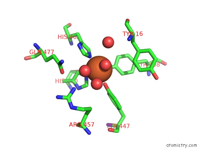
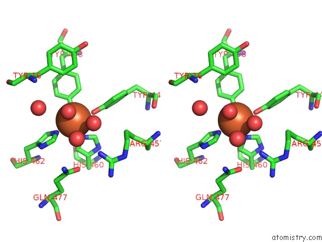
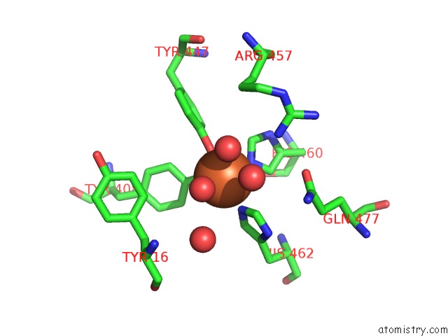
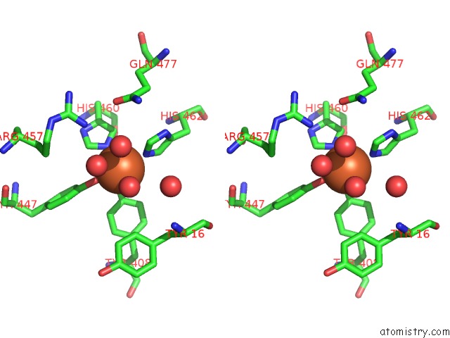
Iron binding site 1 out of 6 in 2pcd
Go back to
Iron binding site 1 out
of 6 in the Structure of Protocatechuate 3,4-Dioxygenase From Pseudomonas Aeruginosa at 2.15 Angstroms Resolution
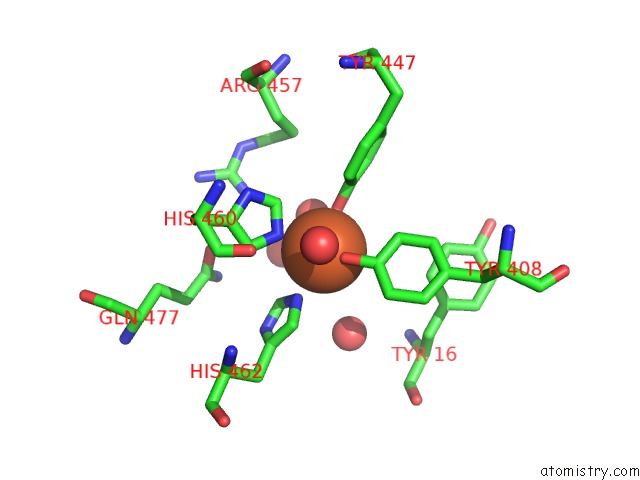
Mono view
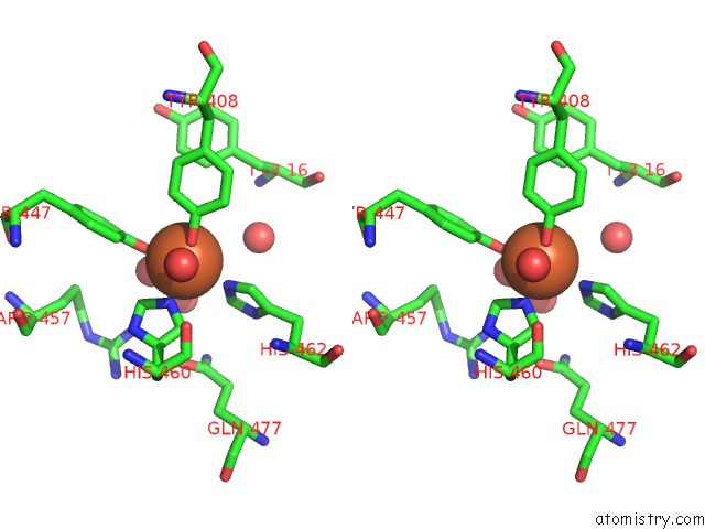
Stereo pair view
Mono view
Stereo pair view
A full contact list of Iron with other atoms in the Fe binding
site number 1 of Structure of Protocatechuate 3,4-Dioxygenase From Pseudomonas Aeruginosa at 2.15 Angstroms Resolution within 5.0Å range:
|
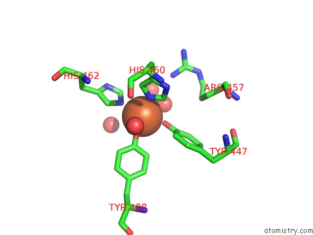
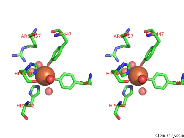
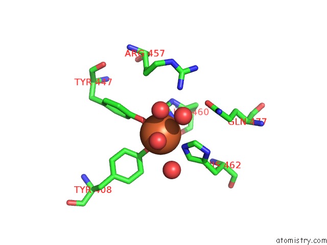
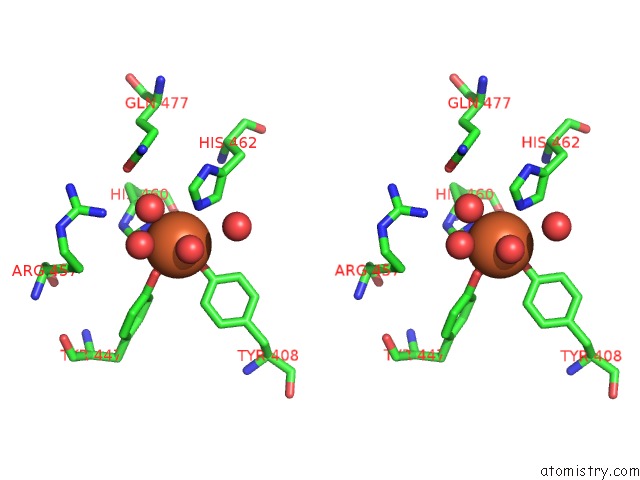
Iron binding site 2 out of 6 in 2pcd
Go back to
Iron binding site 2 out
of 6 in the Structure of Protocatechuate 3,4-Dioxygenase From Pseudomonas Aeruginosa at 2.15 Angstroms Resolution
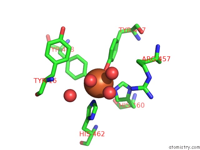
Mono view
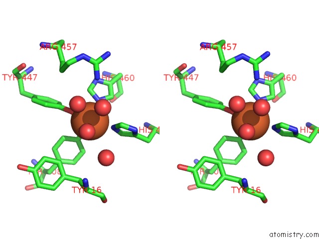
Stereo pair view
Mono view
Stereo pair view
A full contact list of Iron with other atoms in the Fe binding
site number 2 of Structure of Protocatechuate 3,4-Dioxygenase From Pseudomonas Aeruginosa at 2.15 Angstroms Resolution within 5.0Å range:
|
Iron binding site 3 out of 6 in 2pcd
Go back to
Iron binding site 3 out
of 6 in the Structure of Protocatechuate 3,4-Dioxygenase From Pseudomonas Aeruginosa at 2.15 Angstroms Resolution
Mono view
Stereo pair view
Mono view
Stereo pair view
A full contact list of Iron with other atoms in the Fe binding
site number 3 of Structure of Protocatechuate 3,4-Dioxygenase From Pseudomonas Aeruginosa at 2.15 Angstroms Resolution within 5.0Å range:
|
Iron binding site 4 out of 6 in 2pcd
Go back to
Iron binding site 4 out
of 6 in the Structure of Protocatechuate 3,4-Dioxygenase From Pseudomonas Aeruginosa at 2.15 Angstroms Resolution
Mono view
Stereo pair view
Mono view
Stereo pair view
A full contact list of Iron with other atoms in the Fe binding
site number 4 of Structure of Protocatechuate 3,4-Dioxygenase From Pseudomonas Aeruginosa at 2.15 Angstroms Resolution within 5.0Å range:
|
Iron binding site 5 out of 6 in 2pcd
Go back to
Iron binding site 5 out
of 6 in the Structure of Protocatechuate 3,4-Dioxygenase From Pseudomonas Aeruginosa at 2.15 Angstroms Resolution
Mono view
Stereo pair view
Mono view
Stereo pair view
A full contact list of Iron with other atoms in the Fe binding
site number 5 of Structure of Protocatechuate 3,4-Dioxygenase From Pseudomonas Aeruginosa at 2.15 Angstroms Resolution within 5.0Å range:
|
Iron binding site 6 out of 6 in 2pcd
Go back to
Iron binding site 6 out
of 6 in the Structure of Protocatechuate 3,4-Dioxygenase From Pseudomonas Aeruginosa at 2.15 Angstroms Resolution
Mono view
Stereo pair view
Mono view
Stereo pair view
A full contact list of Iron with other atoms in the Fe binding
site number 6 of Structure of Protocatechuate 3,4-Dioxygenase From Pseudomonas Aeruginosa at 2.15 Angstroms Resolution within 5.0Å range:
|
Reference:
D.H.Ohlendorf,
A.M.Orville,
J.D.Lipscomb.
Structure of Protocatechuate 3,4-Dioxygenase From Pseudomonas Aeruginosa at 2.15 A Resolution. J.Mol.Biol. V. 244 586 1994.
ISSN: ISSN 0022-2836
PubMed: 7990141
DOI: 10.1006/JMBI.1994.1754
Page generated: Thu Jul 17 03:24:12 2025
ISSN: ISSN 0022-2836
PubMed: 7990141
DOI: 10.1006/JMBI.1994.1754
Last articles
Fe in 2YXOFe in 2YRS
Fe in 2YXC
Fe in 2YNM
Fe in 2YVJ
Fe in 2YP1
Fe in 2YU2
Fe in 2YU1
Fe in 2YQB
Fe in 2YOO