Iron »
PDB 2pgh-2q9u »
2pms »
Iron in PDB 2pms: Crystal Structure of the Complex of Human Lactoferrin N-Lobe and Lactoferrin-Binding Domain of Pneumococcal Surface Protein A
Protein crystallography data
The structure of Crystal Structure of the Complex of Human Lactoferrin N-Lobe and Lactoferrin-Binding Domain of Pneumococcal Surface Protein A, PDB code: 2pms
was solved by
D.Chattopadhyay,
O.Senkovich,
W.J.Cook,
with X-Ray Crystallography technique. A brief refinement statistics is given in the table below:
| Resolution Low / High (Å) | 15.00 / 2.91 |
| Space group | P 32 |
| Cell size a, b, c (Å), α, β, γ (°) | 130.180, 130.180, 80.800, 90.00, 90.00, 120.00 |
| R / Rfree (%) | 20.3 / 24.9 |
Other elements in 2pms:
The structure of Crystal Structure of the Complex of Human Lactoferrin N-Lobe and Lactoferrin-Binding Domain of Pneumococcal Surface Protein A also contains other interesting chemical elements:
| Zinc | (Zn) | 2 atoms |
Iron Binding Sites:
The binding sites of Iron atom in the Crystal Structure of the Complex of Human Lactoferrin N-Lobe and Lactoferrin-Binding Domain of Pneumococcal Surface Protein A
(pdb code 2pms). This binding sites where shown within
5.0 Angstroms radius around Iron atom.
In total 2 binding sites of Iron where determined in the Crystal Structure of the Complex of Human Lactoferrin N-Lobe and Lactoferrin-Binding Domain of Pneumococcal Surface Protein A, PDB code: 2pms:
Jump to Iron binding site number: 1; 2;
In total 2 binding sites of Iron where determined in the Crystal Structure of the Complex of Human Lactoferrin N-Lobe and Lactoferrin-Binding Domain of Pneumococcal Surface Protein A, PDB code: 2pms:
Jump to Iron binding site number: 1; 2;
Iron binding site 1 out of 2 in 2pms
Go back to
Iron binding site 1 out
of 2 in the Crystal Structure of the Complex of Human Lactoferrin N-Lobe and Lactoferrin-Binding Domain of Pneumococcal Surface Protein A
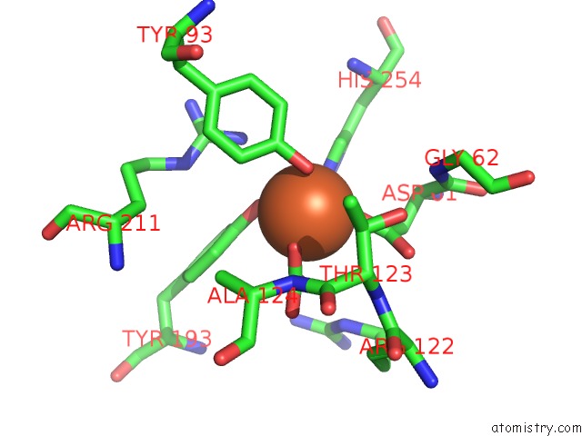
Mono view
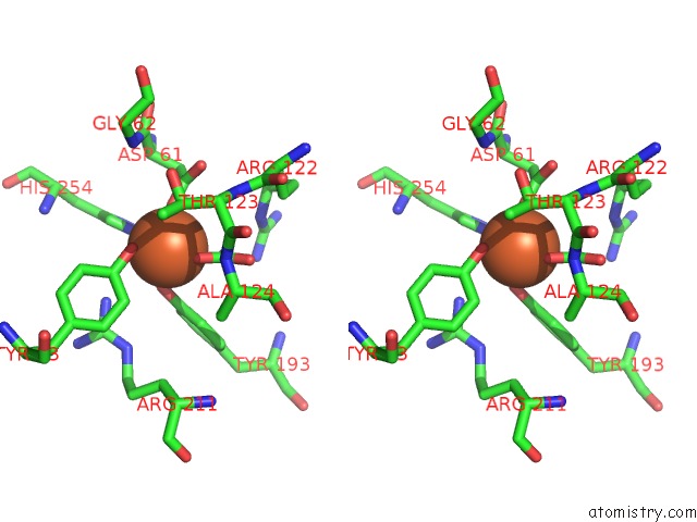
Stereo pair view
Mono view
Stereo pair view
A full contact list of Iron with other atoms in the Fe binding
site number 1 of Crystal Structure of the Complex of Human Lactoferrin N-Lobe and Lactoferrin-Binding Domain of Pneumococcal Surface Protein A within 5.0Å range:
|
Iron binding site 2 out of 2 in 2pms
Go back to
Iron binding site 2 out
of 2 in the Crystal Structure of the Complex of Human Lactoferrin N-Lobe and Lactoferrin-Binding Domain of Pneumococcal Surface Protein A
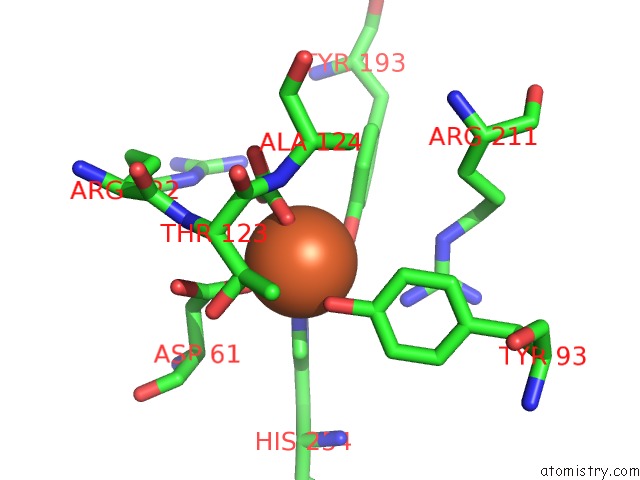
Mono view
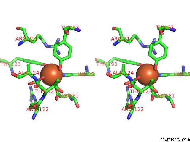
Stereo pair view
Mono view
Stereo pair view
A full contact list of Iron with other atoms in the Fe binding
site number 2 of Crystal Structure of the Complex of Human Lactoferrin N-Lobe and Lactoferrin-Binding Domain of Pneumococcal Surface Protein A within 5.0Å range:
|
Reference:
O.Senkovich,
W.J.Cook,
S.Mirza,
S.K.Hollingshead,
I.I.Protasevich,
D.E.Briles,
D.Chattopadhyay.
Structure of A Complex of Human Lactoferrin N-Lobe with Pneumococcal Surface Protein A Provides Insight Into Microbial Defense Mechanism. J.Mol.Biol. V. 370 701 2007.
ISSN: ISSN 0022-2836
PubMed: 17543335
DOI: 10.1016/J.JMB.2007.04.075
Page generated: Sun Aug 4 01:27:04 2024
ISSN: ISSN 0022-2836
PubMed: 17543335
DOI: 10.1016/J.JMB.2007.04.075
Last articles
F in 7RBTF in 7R9Y
F in 7R9N
F in 7R9Z
F in 7R9P
F in 7R9T
F in 7R9V
F in 7R9C
F in 7QZ7
F in 7R9F