Iron »
PDB 2pgh-2q9u »
2pt2 »
Iron in PDB 2pt2: Structure of FUTA1 with Iron(II)
Protein crystallography data
The structure of Structure of FUTA1 with Iron(II), PDB code: 2pt2
was solved by
N.M.Koropatkin,
A.M.Randich,
M.Bhattachryya-Pakrasi,
H.B.Pakrasi,
T.J.Smith,
with X-Ray Crystallography technique. A brief refinement statistics is given in the table below:
| Resolution Low / High (Å) | 50.00 / 2.00 |
| Space group | C 1 2 1 |
| Cell size a, b, c (Å), α, β, γ (°) | 98.307, 65.703, 50.255, 90.00, 102.43, 90.00 |
| R / Rfree (%) | 17.5 / 21.2 |
Iron Binding Sites:
The binding sites of Iron atom in the Structure of FUTA1 with Iron(II)
(pdb code 2pt2). This binding sites where shown within
5.0 Angstroms radius around Iron atom.
In total only one binding site of Iron was determined in the Structure of FUTA1 with Iron(II), PDB code: 2pt2:
In total only one binding site of Iron was determined in the Structure of FUTA1 with Iron(II), PDB code: 2pt2:
Iron binding site 1 out of 1 in 2pt2
Go back to
Iron binding site 1 out
of 1 in the Structure of FUTA1 with Iron(II)
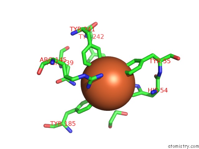
Mono view
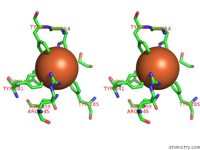
Stereo pair view
Mono view
Stereo pair view
A full contact list of Iron with other atoms in the Fe binding
site number 1 of Structure of FUTA1 with Iron(II) within 5.0Å range:
|
Reference:
N.Koropatkin,
A.M.Randich,
M.Bhattacharyya-Pakrasi,
H.B.Pakrasi,
T.J.Smith.
The Structure of the Iron-Binding Protein, FUTA1, From Synechocystis 6803. J.Biol.Chem. V. 282 27468 2007.
ISSN: ISSN 0021-9258
PubMed: 17626019
DOI: 10.1074/JBC.M704136200
Page generated: Thu Jul 17 03:30:55 2025
ISSN: ISSN 0021-9258
PubMed: 17626019
DOI: 10.1074/JBC.M704136200
Last articles
Fe in 2YXOFe in 2YRS
Fe in 2YXC
Fe in 2YNM
Fe in 2YVJ
Fe in 2YP1
Fe in 2YU2
Fe in 2YU1
Fe in 2YQB
Fe in 2YOO