Iron »
PDB 2pgh-2q9u »
2pxj »
Iron in PDB 2pxj: The Complex Structure of JMJD2A and Monomethylated H3K36 Peptide
Protein crystallography data
The structure of The Complex Structure of JMJD2A and Monomethylated H3K36 Peptide, PDB code: 2pxj
was solved by
Z.Chen,
J.Zang,
J.Kappler,
X.Hong,
F.Crawford,
G.Zhang,
with X-Ray Crystallography technique. A brief refinement statistics is given in the table below:
| Resolution Low / High (Å) | 45.56 / 2.00 |
| Space group | P 21 21 2 |
| Cell size a, b, c (Å), α, β, γ (°) | 100.863, 150.172, 57.317, 90.00, 90.00, 90.00 |
| R / Rfree (%) | 25.6 / 27.6 |
Other elements in 2pxj:
The structure of The Complex Structure of JMJD2A and Monomethylated H3K36 Peptide also contains other interesting chemical elements:
| Zinc | (Zn) | 2 atoms |
Iron Binding Sites:
The binding sites of Iron atom in the The Complex Structure of JMJD2A and Monomethylated H3K36 Peptide
(pdb code 2pxj). This binding sites where shown within
5.0 Angstroms radius around Iron atom.
In total 2 binding sites of Iron where determined in the The Complex Structure of JMJD2A and Monomethylated H3K36 Peptide, PDB code: 2pxj:
Jump to Iron binding site number: 1; 2;
In total 2 binding sites of Iron where determined in the The Complex Structure of JMJD2A and Monomethylated H3K36 Peptide, PDB code: 2pxj:
Jump to Iron binding site number: 1; 2;
Iron binding site 1 out of 2 in 2pxj
Go back to
Iron binding site 1 out
of 2 in the The Complex Structure of JMJD2A and Monomethylated H3K36 Peptide
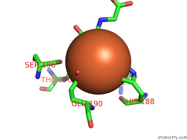
Mono view
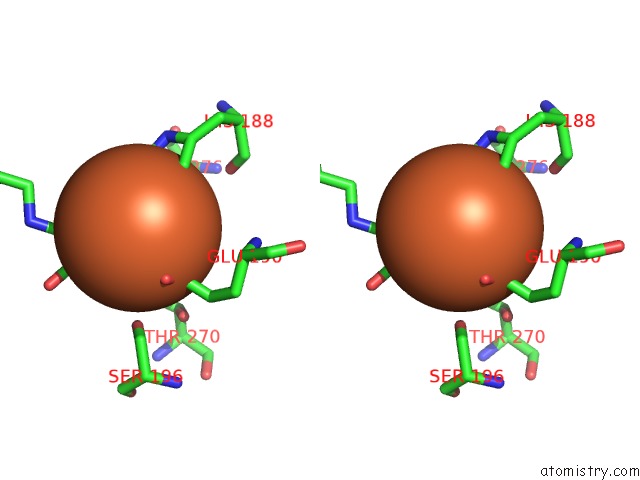
Stereo pair view
Mono view
Stereo pair view
A full contact list of Iron with other atoms in the Fe binding
site number 1 of The Complex Structure of JMJD2A and Monomethylated H3K36 Peptide within 5.0Å range:
|
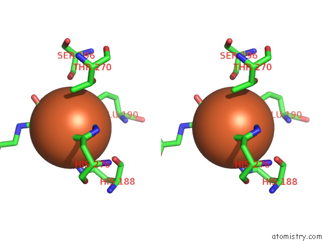
Iron binding site 2 out of 2 in 2pxj
Go back to
Iron binding site 2 out
of 2 in the The Complex Structure of JMJD2A and Monomethylated H3K36 Peptide
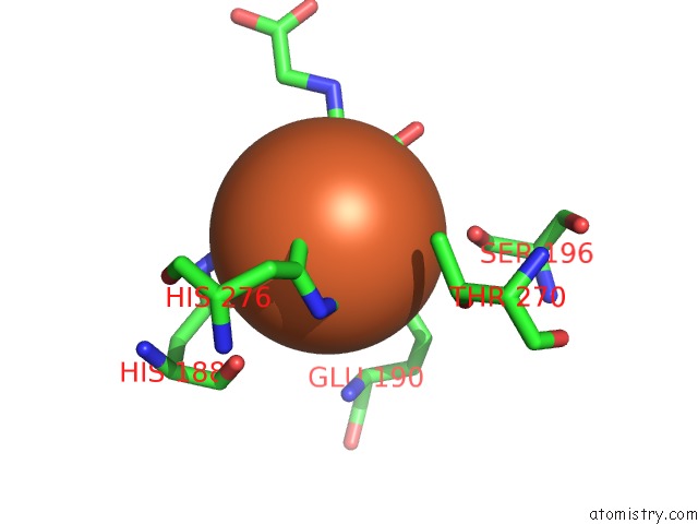
Mono view
Stereo pair view
Mono view
Stereo pair view
A full contact list of Iron with other atoms in the Fe binding
site number 2 of The Complex Structure of JMJD2A and Monomethylated H3K36 Peptide within 5.0Å range:
|
Reference:
Z.Chen,
J.Zang,
J.Kappler,
X.Hong,
F.Crawford,
Q.Wang,
F.Lan,
C.Jiang,
J.Whetstine,
S.Dai,
K.Hansen,
Y.Shi,
G.Zhang.
Structural Basis of the Recognition of A Methylated Histone Tail By JMJD2A Proc.Natl.Acad.Sci.Usa V. 104 10818 2007.
ISSN: ISSN 0027-8424
PubMed: 17567753
DOI: 10.1073/PNAS.0704525104
Page generated: Sun Aug 4 01:33:59 2024
ISSN: ISSN 0027-8424
PubMed: 17567753
DOI: 10.1073/PNAS.0704525104
Last articles
Zn in 9MJ5Zn in 9HNW
Zn in 9G0L
Zn in 9FNE
Zn in 9DZN
Zn in 9E0I
Zn in 9D32
Zn in 9DAK
Zn in 8ZXC
Zn in 8ZUF