Iron »
PDB 2pgh-2q9u »
2q09 »
Iron in PDB 2q09: Crystal Structure of Imidazolonepropionase From Environmental Sample with Bound Inhibitor 3-(2,5-Dioxo-Imidazolidin-4-Yl)-Propionic Acid
Enzymatic activity of Crystal Structure of Imidazolonepropionase From Environmental Sample with Bound Inhibitor 3-(2,5-Dioxo-Imidazolidin-4-Yl)-Propionic Acid
All present enzymatic activity of Crystal Structure of Imidazolonepropionase From Environmental Sample with Bound Inhibitor 3-(2,5-Dioxo-Imidazolidin-4-Yl)-Propionic Acid:
3.5.2.7;
3.5.2.7;
Protein crystallography data
The structure of Crystal Structure of Imidazolonepropionase From Environmental Sample with Bound Inhibitor 3-(2,5-Dioxo-Imidazolidin-4-Yl)-Propionic Acid, PDB code: 2q09
was solved by
R.Tyagi,
S.Eswaramoorthy,
S.K.Burley,
S.Swaminathan,
New York Sgxresearch Center For Structural Genomics (Nysgxrc),
with X-Ray Crystallography technique. A brief refinement statistics is given in the table below:
| Resolution Low / High (Å) | 31.39 / 1.97 |
| Space group | P 32 2 1 |
| Cell size a, b, c (Å), α, β, γ (°) | 95.910, 95.910, 115.473, 90.00, 90.00, 120.00 |
| R / Rfree (%) | 19.4 / 21.4 |
Iron Binding Sites:
The binding sites of Iron atom in the Crystal Structure of Imidazolonepropionase From Environmental Sample with Bound Inhibitor 3-(2,5-Dioxo-Imidazolidin-4-Yl)-Propionic Acid
(pdb code 2q09). This binding sites where shown within
5.0 Angstroms radius around Iron atom.
In total only one binding site of Iron was determined in the Crystal Structure of Imidazolonepropionase From Environmental Sample with Bound Inhibitor 3-(2,5-Dioxo-Imidazolidin-4-Yl)-Propionic Acid, PDB code: 2q09:
In total only one binding site of Iron was determined in the Crystal Structure of Imidazolonepropionase From Environmental Sample with Bound Inhibitor 3-(2,5-Dioxo-Imidazolidin-4-Yl)-Propionic Acid, PDB code: 2q09:
Iron binding site 1 out of 1 in 2q09
Go back to
Iron binding site 1 out
of 1 in the Crystal Structure of Imidazolonepropionase From Environmental Sample with Bound Inhibitor 3-(2,5-Dioxo-Imidazolidin-4-Yl)-Propionic Acid
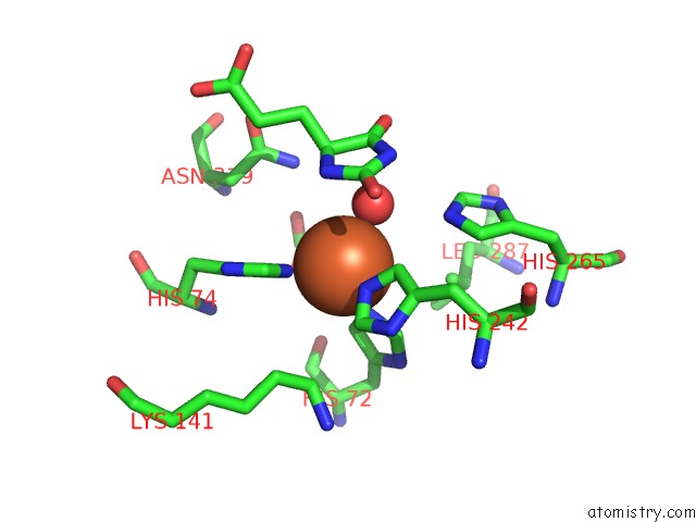
Mono view
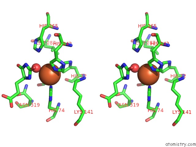
Stereo pair view
Mono view
Stereo pair view
A full contact list of Iron with other atoms in the Fe binding
site number 1 of Crystal Structure of Imidazolonepropionase From Environmental Sample with Bound Inhibitor 3-(2,5-Dioxo-Imidazolidin-4-Yl)-Propionic Acid within 5.0Å range:
|
Reference:
R.Tyagi,
S.Eswaramoorthy,
S.K.Burley,
F.M.Raushel,
S.Swaminathan.
A Common Catalytic Mechanism For Proteins of the Huti Family. Biochemistry V. 47 5608 2008.
ISSN: ISSN 0006-2960
PubMed: 18442260
DOI: 10.1021/BI800180G
Page generated: Sun Aug 4 01:34:44 2024
ISSN: ISSN 0006-2960
PubMed: 18442260
DOI: 10.1021/BI800180G
Last articles
F in 7NTHF in 7NTI
F in 7NPC
F in 7NRG
F in 7NR5
F in 7NQS
F in 7NOS
F in 7NP5
F in 7NDV
F in 7NP6