Iron »
PDB 2pgh-2q9u »
2q8j »
Iron in PDB 2q8j: Crystal Structure of the Complex of C-Lobe of Bovine Lactoferrin with Mannitol and Mannose at 2.7 A Resolution
Protein crystallography data
The structure of Crystal Structure of the Complex of C-Lobe of Bovine Lactoferrin with Mannitol and Mannose at 2.7 A Resolution, PDB code: 2q8j
was solved by
R.Mir,
R.Jain,
M.Sinha,
N.Singh,
S.Sharma,
P.Kaur,
A.Bhushan,
T.P.Singh,
with X-Ray Crystallography technique. A brief refinement statistics is given in the table below:
| Resolution Low / High (Å) | 20.00 / 2.71 |
| Space group | P 1 21 1 |
| Cell size a, b, c (Å), α, β, γ (°) | 63.400, 50.300, 65.900, 90.00, 107.80, 90.00 |
| R / Rfree (%) | 19.5 / 24 |
Other elements in 2q8j:
The structure of Crystal Structure of the Complex of C-Lobe of Bovine Lactoferrin with Mannitol and Mannose at 2.7 A Resolution also contains other interesting chemical elements:
| Zinc | (Zn) | 2 atoms |
Iron Binding Sites:
The binding sites of Iron atom in the Crystal Structure of the Complex of C-Lobe of Bovine Lactoferrin with Mannitol and Mannose at 2.7 A Resolution
(pdb code 2q8j). This binding sites where shown within
5.0 Angstroms radius around Iron atom.
In total only one binding site of Iron was determined in the Crystal Structure of the Complex of C-Lobe of Bovine Lactoferrin with Mannitol and Mannose at 2.7 A Resolution, PDB code: 2q8j:
In total only one binding site of Iron was determined in the Crystal Structure of the Complex of C-Lobe of Bovine Lactoferrin with Mannitol and Mannose at 2.7 A Resolution, PDB code: 2q8j:
Iron binding site 1 out of 1 in 2q8j
Go back to
Iron binding site 1 out
of 1 in the Crystal Structure of the Complex of C-Lobe of Bovine Lactoferrin with Mannitol and Mannose at 2.7 A Resolution
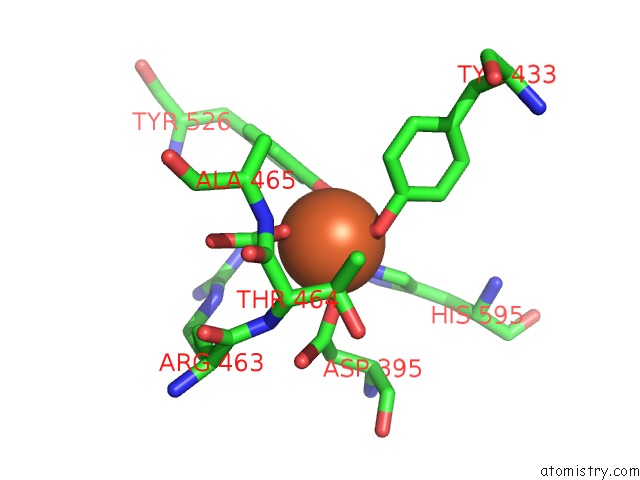
Mono view
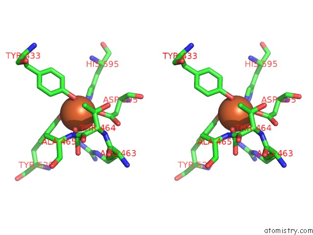
Stereo pair view
Mono view
Stereo pair view
A full contact list of Iron with other atoms in the Fe binding
site number 1 of Crystal Structure of the Complex of C-Lobe of Bovine Lactoferrin with Mannitol and Mannose at 2.7 A Resolution within 5.0Å range:
|
Reference:
R.Mir,
R.Jain,
M.Sinha,
N.Singh,
S.Sharma,
P.Kaur,
A.Bhushan,
T.P.Singh.
Crystal Structure of the Complex of C-Lobe of Bovine Lactoferrin with Mannitol and Mannose at 2.7 A Resolution To Be Published.
Page generated: Sun Aug 4 01:37:52 2024
Last articles
Cl in 8BSRCl in 8BRN
Cl in 8BS0
Cl in 8BRZ
Cl in 8BRY
Cl in 8BQQ
Cl in 8BQT
Cl in 8BRE
Cl in 8BQH
Cl in 8BQP