Iron »
PDB 2qbl-2r1l »
2qd0 »
Iron in PDB 2qd0: Crystal Structure of Mitoneet
Protein crystallography data
The structure of Crystal Structure of Mitoneet, PDB code: 2qd0
was solved by
J.Lin,
T.Zhou,
K.Ye,
J.Wang,
with X-Ray Crystallography technique. A brief refinement statistics is given in the table below:
| Resolution Low / High (Å) | 21.92 / 1.81 |
| Space group | P 21 21 21 |
| Cell size a, b, c (Å), α, β, γ (°) | 43.838, 59.139, 65.971, 90.00, 90.00, 90.00 |
| R / Rfree (%) | 16.4 / 19.3 |
Iron Binding Sites:
The binding sites of Iron atom in the Crystal Structure of Mitoneet
(pdb code 2qd0). This binding sites where shown within
5.0 Angstroms radius around Iron atom.
In total 4 binding sites of Iron where determined in the Crystal Structure of Mitoneet, PDB code: 2qd0:
Jump to Iron binding site number: 1; 2; 3; 4;
In total 4 binding sites of Iron where determined in the Crystal Structure of Mitoneet, PDB code: 2qd0:
Jump to Iron binding site number: 1; 2; 3; 4;
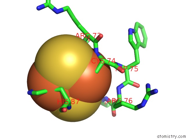
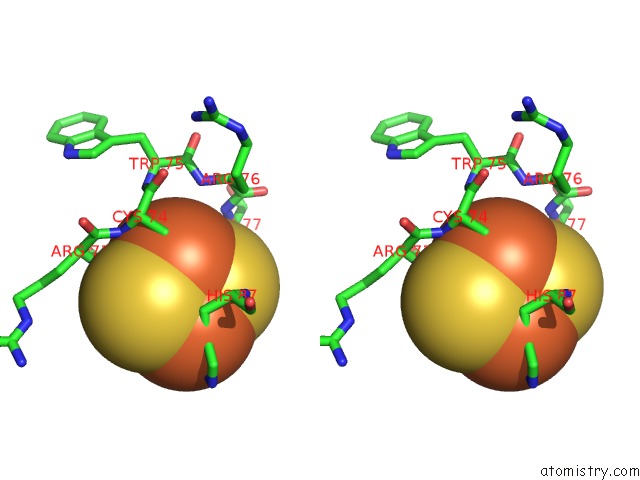
Iron binding site 1 out of 4 in 2qd0
Go back to
Iron binding site 1 out
of 4 in the Crystal Structure of Mitoneet
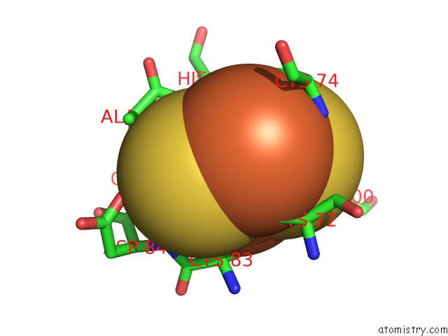
Mono view
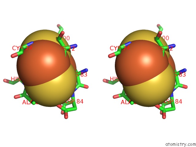
Stereo pair view
Mono view
Stereo pair view
A full contact list of Iron with other atoms in the Fe binding
site number 1 of Crystal Structure of Mitoneet within 5.0Å range:
|
Iron binding site 2 out of 4 in 2qd0
Go back to
Iron binding site 2 out
of 4 in the Crystal Structure of Mitoneet
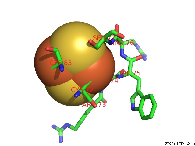
Mono view
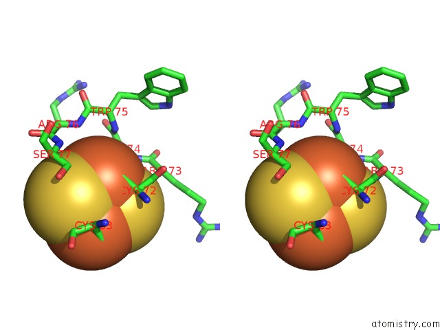
Stereo pair view
Mono view
Stereo pair view
A full contact list of Iron with other atoms in the Fe binding
site number 2 of Crystal Structure of Mitoneet within 5.0Å range:
|
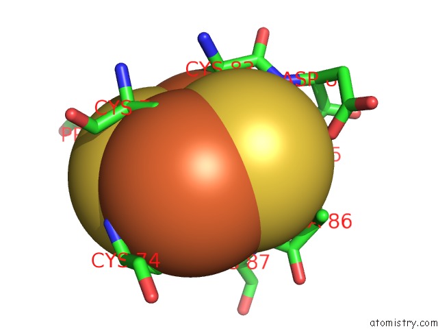
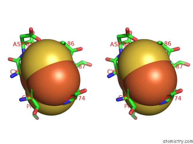
Iron binding site 3 out of 4 in 2qd0
Go back to
Iron binding site 3 out
of 4 in the Crystal Structure of Mitoneet
Mono view
Stereo pair view
Mono view
Stereo pair view
A full contact list of Iron with other atoms in the Fe binding
site number 3 of Crystal Structure of Mitoneet within 5.0Å range:
|
Iron binding site 4 out of 4 in 2qd0
Go back to
Iron binding site 4 out
of 4 in the Crystal Structure of Mitoneet
Mono view
Stereo pair view
Mono view
Stereo pair view
A full contact list of Iron with other atoms in the Fe binding
site number 4 of Crystal Structure of Mitoneet within 5.0Å range:
|
Reference:
J.Lin,
T.Zhou,
K.Ye,
J.Wang.
Crystal Structure of Human Mitoneet Reveals Distinct Groups of Iron Sulfur Proteins. Proc.Natl.Acad.Sci.Usa V. 104 14640 2007.
ISSN: ISSN 0027-8424
PubMed: 17766439
DOI: 10.1073/PNAS.0702426104
Page generated: Sun Aug 4 01:41:14 2024
ISSN: ISSN 0027-8424
PubMed: 17766439
DOI: 10.1073/PNAS.0702426104
Last articles
Cl in 5SYICl in 5SYH
Cl in 5SXX
Cl in 5SXT
Cl in 5SXU
Cl in 5SXS
Cl in 5SXR
Cl in 5SXQ
Cl in 5SX3
Cl in 5SWR