Iron »
PDB 2qbl-2r1l »
2qdy »
Iron in PDB 2qdy: Crystal Structure of Fe-Type Nhase From Rhodococcus Erythropolis AJ270
Enzymatic activity of Crystal Structure of Fe-Type Nhase From Rhodococcus Erythropolis AJ270
All present enzymatic activity of Crystal Structure of Fe-Type Nhase From Rhodococcus Erythropolis AJ270:
4.2.1.84;
4.2.1.84;
Protein crystallography data
The structure of Crystal Structure of Fe-Type Nhase From Rhodococcus Erythropolis AJ270, PDB code: 2qdy
was solved by
L.Song,
J.Shi,
Z.Xue,
M.-X.Wang,
S.Qian,
with X-Ray Crystallography technique. A brief refinement statistics is given in the table below:
| Resolution Low / High (Å) | 35.30 / 1.30 |
| Space group | C 1 2 1 |
| Cell size a, b, c (Å), α, β, γ (°) | 114.064, 60.068, 81.761, 90.00, 125.15, 90.00 |
| R / Rfree (%) | 12.9 / 15.7 |
Other elements in 2qdy:
The structure of Crystal Structure of Fe-Type Nhase From Rhodococcus Erythropolis AJ270 also contains other interesting chemical elements:
| Magnesium | (Mg) | 3 atoms |
| Chlorine | (Cl) | 4 atoms |
Iron Binding Sites:
The binding sites of Iron atom in the Crystal Structure of Fe-Type Nhase From Rhodococcus Erythropolis AJ270
(pdb code 2qdy). This binding sites where shown within
5.0 Angstroms radius around Iron atom.
In total only one binding site of Iron was determined in the Crystal Structure of Fe-Type Nhase From Rhodococcus Erythropolis AJ270, PDB code: 2qdy:
In total only one binding site of Iron was determined in the Crystal Structure of Fe-Type Nhase From Rhodococcus Erythropolis AJ270, PDB code: 2qdy:
Iron binding site 1 out of 1 in 2qdy
Go back to
Iron binding site 1 out
of 1 in the Crystal Structure of Fe-Type Nhase From Rhodococcus Erythropolis AJ270
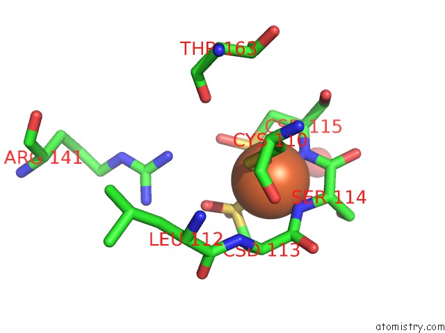
Mono view
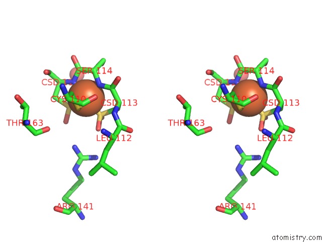
Stereo pair view
Mono view
Stereo pair view
A full contact list of Iron with other atoms in the Fe binding
site number 1 of Crystal Structure of Fe-Type Nhase From Rhodococcus Erythropolis AJ270 within 5.0Å range:
|
Reference:
L.Song,
M.Wang,
J.Shi,
Z.Xue,
M.-X.Wang,
S.Qian.
High Resolution X-Ray Molecular Structure of the Nitrile Hydratase From Rhodococcus Erythropolis AJ270 Reveals Posttranslational Oxidation of Two Cysteines Into Sulfinic Acids and A Novel Biocatalytic Nitrile Hydration Mechanism Biochem.Biophys.Res.Commun. V. 362 319 2007.
ISSN: ISSN 0006-291X
PubMed: 17716629
DOI: 10.1016/J.BBRC.2007.07.184
Page generated: Sun Aug 4 01:42:41 2024
ISSN: ISSN 0006-291X
PubMed: 17716629
DOI: 10.1016/J.BBRC.2007.07.184
Last articles
Zn in 9MJ5Zn in 9HNW
Zn in 9G0L
Zn in 9FNE
Zn in 9DZN
Zn in 9E0I
Zn in 9D32
Zn in 9DAK
Zn in 8ZXC
Zn in 8ZUF