Iron »
PDB 2qbl-2r1l »
2qfk »
Iron in PDB 2qfk: X-Ray Crystal Structure Analysis of the Binding Site in the Ferric and Oxyferrous Forms of the Recombinant Heme Dehaloperoxidase Cloned From Amphitrite Ornata
Protein crystallography data
The structure of X-Ray Crystal Structure Analysis of the Binding Site in the Ferric and Oxyferrous Forms of the Recombinant Heme Dehaloperoxidase Cloned From Amphitrite Ornata, PDB code: 2qfk
was solved by
V.S.De Serrano,
Z.Chen,
M.F.Davis,
S.Franzen,
with X-Ray Crystallography technique. A brief refinement statistics is given in the table below:
| Resolution Low / High (Å) | 25.62 / 1.62 |
| Space group | P 21 21 21 |
| Cell size a, b, c (Å), α, β, γ (°) | 60.686, 67.572, 67.622, 90.00, 90.00, 90.00 |
| R / Rfree (%) | 19.1 / 23.9 |
Iron Binding Sites:
The binding sites of Iron atom in the X-Ray Crystal Structure Analysis of the Binding Site in the Ferric and Oxyferrous Forms of the Recombinant Heme Dehaloperoxidase Cloned From Amphitrite Ornata
(pdb code 2qfk). This binding sites where shown within
5.0 Angstroms radius around Iron atom.
In total 2 binding sites of Iron where determined in the X-Ray Crystal Structure Analysis of the Binding Site in the Ferric and Oxyferrous Forms of the Recombinant Heme Dehaloperoxidase Cloned From Amphitrite Ornata, PDB code: 2qfk:
Jump to Iron binding site number: 1; 2;
In total 2 binding sites of Iron where determined in the X-Ray Crystal Structure Analysis of the Binding Site in the Ferric and Oxyferrous Forms of the Recombinant Heme Dehaloperoxidase Cloned From Amphitrite Ornata, PDB code: 2qfk:
Jump to Iron binding site number: 1; 2;
Iron binding site 1 out of 2 in 2qfk
Go back to
Iron binding site 1 out
of 2 in the X-Ray Crystal Structure Analysis of the Binding Site in the Ferric and Oxyferrous Forms of the Recombinant Heme Dehaloperoxidase Cloned From Amphitrite Ornata
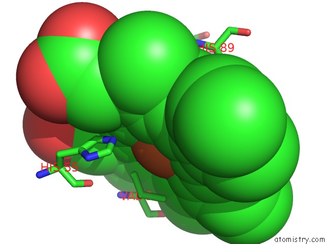
Mono view
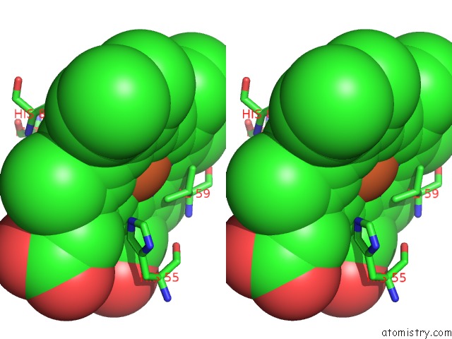
Stereo pair view
Mono view
Stereo pair view
A full contact list of Iron with other atoms in the Fe binding
site number 1 of X-Ray Crystal Structure Analysis of the Binding Site in the Ferric and Oxyferrous Forms of the Recombinant Heme Dehaloperoxidase Cloned From Amphitrite Ornata within 5.0Å range:
|
Iron binding site 2 out of 2 in 2qfk
Go back to
Iron binding site 2 out
of 2 in the X-Ray Crystal Structure Analysis of the Binding Site in the Ferric and Oxyferrous Forms of the Recombinant Heme Dehaloperoxidase Cloned From Amphitrite Ornata
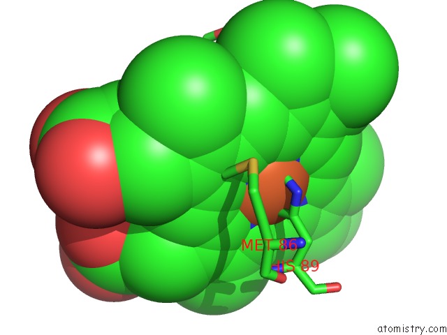
Mono view
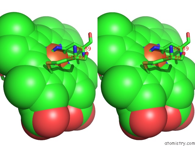
Stereo pair view
Mono view
Stereo pair view
A full contact list of Iron with other atoms in the Fe binding
site number 2 of X-Ray Crystal Structure Analysis of the Binding Site in the Ferric and Oxyferrous Forms of the Recombinant Heme Dehaloperoxidase Cloned From Amphitrite Ornata within 5.0Å range:
|
Reference:
V.S.De Serrano,
Z.Chen,
M.F.Davis,
S.Franzen.
X-Ray Crystal Structural Analysis of the Binding Site in the Ferric and Oxyferrous Forms of the Recombinant Heme Dehaloperoxidase Cloned From Amphitrite Ornata Acta Crystallogr.,Sect.D V. 63 1094 2007.
ISSN: ISSN 0907-4449
PubMed: 17881827
DOI: 10.1107/S0907444907043417
Page generated: Thu Jul 17 03:40:38 2025
ISSN: ISSN 0907-4449
PubMed: 17881827
DOI: 10.1107/S0907444907043417
Last articles
Fe in 2YXOFe in 2YRS
Fe in 2YXC
Fe in 2YNM
Fe in 2YVJ
Fe in 2YP1
Fe in 2YU2
Fe in 2YU1
Fe in 2YQB
Fe in 2YOO