Iron »
PDB 2qbl-2r1l »
2qjk »
Iron in PDB 2qjk: Crystal Structure Analysis of Mutant Rhodobacter Sphaeroides BC1 with Stigmatellin and Antimycin
Enzymatic activity of Crystal Structure Analysis of Mutant Rhodobacter Sphaeroides BC1 with Stigmatellin and Antimycin
All present enzymatic activity of Crystal Structure Analysis of Mutant Rhodobacter Sphaeroides BC1 with Stigmatellin and Antimycin:
1.10.2.2;
1.10.2.2;
Protein crystallography data
The structure of Crystal Structure Analysis of Mutant Rhodobacter Sphaeroides BC1 with Stigmatellin and Antimycin, PDB code: 2qjk
was solved by
L.Esser,
with X-Ray Crystallography technique. A brief refinement statistics is given in the table below:
| Resolution Low / High (Å) | 18.00 / 3.10 |
| Space group | C 1 2 1 |
| Cell size a, b, c (Å), α, β, γ (°) | 352.291, 147.396, 160.762, 90.00, 104.13, 90.00 |
| R / Rfree (%) | 23.9 / 26.6 |
Other elements in 2qjk:
The structure of Crystal Structure Analysis of Mutant Rhodobacter Sphaeroides BC1 with Stigmatellin and Antimycin also contains other interesting chemical elements:
| Strontium | (Sr) | 6 atoms |
Iron Binding Sites:
Pages:
>>> Page 1 <<< Page 2, Binding sites: 11 - 20; Page 3, Binding sites: 21 - 30;Binding sites:
The binding sites of Iron atom in the Crystal Structure Analysis of Mutant Rhodobacter Sphaeroides BC1 with Stigmatellin and Antimycin (pdb code 2qjk). This binding sites where shown within 5.0 Angstroms radius around Iron atom.In total 30 binding sites of Iron where determined in the Crystal Structure Analysis of Mutant Rhodobacter Sphaeroides BC1 with Stigmatellin and Antimycin, PDB code: 2qjk:
Jump to Iron binding site number: 1; 2; 3; 4; 5; 6; 7; 8; 9; 10;
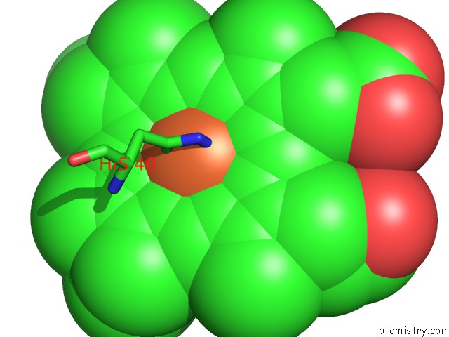
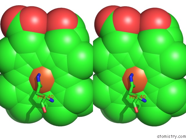
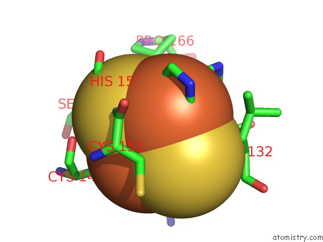
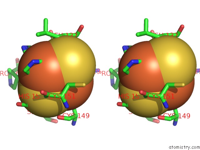
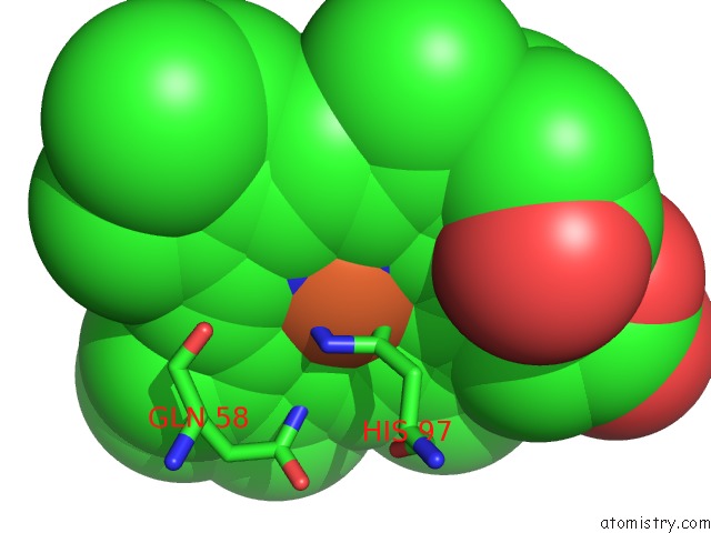
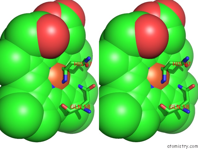
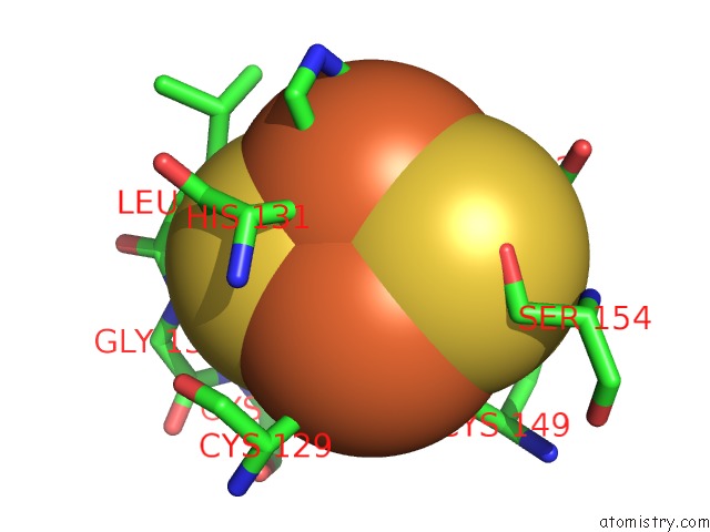
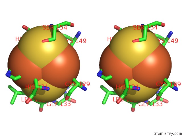
Iron binding site 1 out of 30 in 2qjk
Go back to
Iron binding site 1 out
of 30 in the Crystal Structure Analysis of Mutant Rhodobacter Sphaeroides BC1 with Stigmatellin and Antimycin
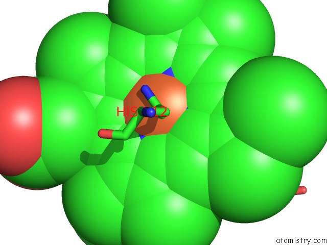
Mono view
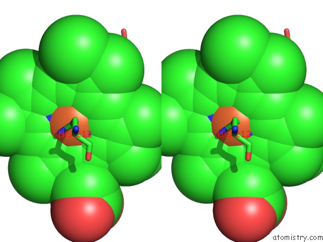
Stereo pair view
Mono view
Stereo pair view
A full contact list of Iron with other atoms in the Fe binding
site number 1 of Crystal Structure Analysis of Mutant Rhodobacter Sphaeroides BC1 with Stigmatellin and Antimycin within 5.0Å range:
|
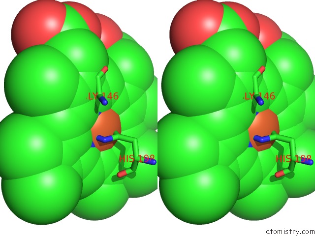
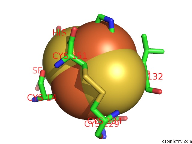
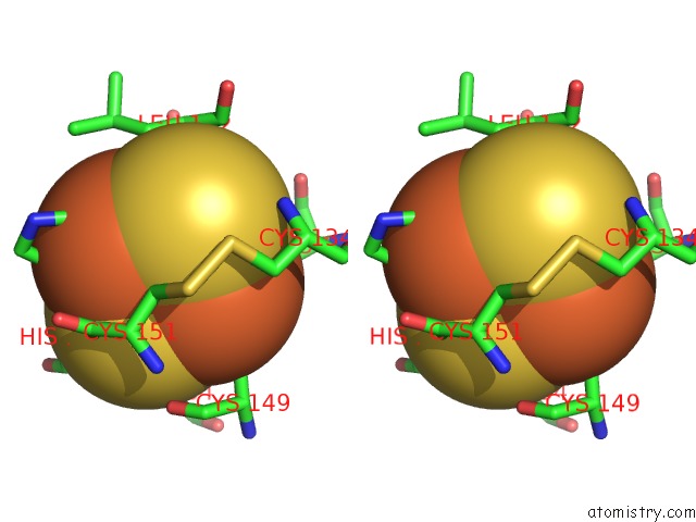
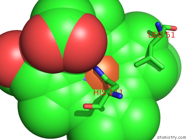
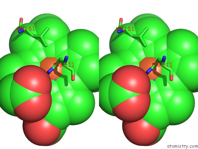
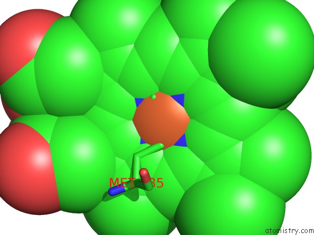
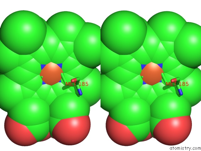
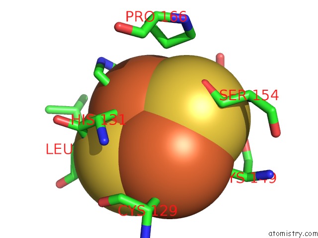
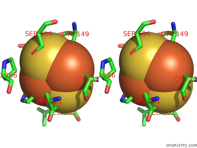
Iron binding site 2 out of 30 in 2qjk
Go back to
Iron binding site 2 out
of 30 in the Crystal Structure Analysis of Mutant Rhodobacter Sphaeroides BC1 with Stigmatellin and Antimycin
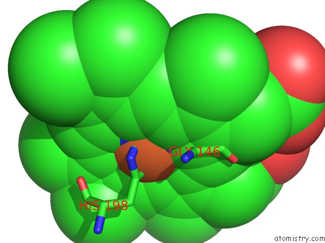
Mono view
Stereo pair view
Mono view
Stereo pair view
A full contact list of Iron with other atoms in the Fe binding
site number 2 of Crystal Structure Analysis of Mutant Rhodobacter Sphaeroides BC1 with Stigmatellin and Antimycin within 5.0Å range:
|
Iron binding site 3 out of 30 in 2qjk
Go back to
Iron binding site 3 out
of 30 in the Crystal Structure Analysis of Mutant Rhodobacter Sphaeroides BC1 with Stigmatellin and Antimycin
Mono view
Stereo pair view
Mono view
Stereo pair view
A full contact list of Iron with other atoms in the Fe binding
site number 3 of Crystal Structure Analysis of Mutant Rhodobacter Sphaeroides BC1 with Stigmatellin and Antimycin within 5.0Å range:
|
Iron binding site 4 out of 30 in 2qjk
Go back to
Iron binding site 4 out
of 30 in the Crystal Structure Analysis of Mutant Rhodobacter Sphaeroides BC1 with Stigmatellin and Antimycin
Mono view
Stereo pair view
Mono view
Stereo pair view
A full contact list of Iron with other atoms in the Fe binding
site number 4 of Crystal Structure Analysis of Mutant Rhodobacter Sphaeroides BC1 with Stigmatellin and Antimycin within 5.0Å range:
|
Iron binding site 5 out of 30 in 2qjk
Go back to
Iron binding site 5 out
of 30 in the Crystal Structure Analysis of Mutant Rhodobacter Sphaeroides BC1 with Stigmatellin and Antimycin
Mono view
Stereo pair view
Mono view
Stereo pair view
A full contact list of Iron with other atoms in the Fe binding
site number 5 of Crystal Structure Analysis of Mutant Rhodobacter Sphaeroides BC1 with Stigmatellin and Antimycin within 5.0Å range:
|
Iron binding site 6 out of 30 in 2qjk
Go back to
Iron binding site 6 out
of 30 in the Crystal Structure Analysis of Mutant Rhodobacter Sphaeroides BC1 with Stigmatellin and Antimycin
Mono view
Stereo pair view
Mono view
Stereo pair view
A full contact list of Iron with other atoms in the Fe binding
site number 6 of Crystal Structure Analysis of Mutant Rhodobacter Sphaeroides BC1 with Stigmatellin and Antimycin within 5.0Å range:
|
Iron binding site 7 out of 30 in 2qjk
Go back to
Iron binding site 7 out
of 30 in the Crystal Structure Analysis of Mutant Rhodobacter Sphaeroides BC1 with Stigmatellin and Antimycin
Mono view
Stereo pair view
Mono view
Stereo pair view
A full contact list of Iron with other atoms in the Fe binding
site number 7 of Crystal Structure Analysis of Mutant Rhodobacter Sphaeroides BC1 with Stigmatellin and Antimycin within 5.0Å range:
|
Iron binding site 8 out of 30 in 2qjk
Go back to
Iron binding site 8 out
of 30 in the Crystal Structure Analysis of Mutant Rhodobacter Sphaeroides BC1 with Stigmatellin and Antimycin
Mono view
Stereo pair view
Mono view
Stereo pair view
A full contact list of Iron with other atoms in the Fe binding
site number 8 of Crystal Structure Analysis of Mutant Rhodobacter Sphaeroides BC1 with Stigmatellin and Antimycin within 5.0Å range:
|
Iron binding site 9 out of 30 in 2qjk
Go back to
Iron binding site 9 out
of 30 in the Crystal Structure Analysis of Mutant Rhodobacter Sphaeroides BC1 with Stigmatellin and Antimycin
Mono view
Stereo pair view
Mono view
Stereo pair view
A full contact list of Iron with other atoms in the Fe binding
site number 9 of Crystal Structure Analysis of Mutant Rhodobacter Sphaeroides BC1 with Stigmatellin and Antimycin within 5.0Å range:
|
Iron binding site 10 out of 30 in 2qjk
Go back to
Iron binding site 10 out
of 30 in the Crystal Structure Analysis of Mutant Rhodobacter Sphaeroides BC1 with Stigmatellin and Antimycin
Mono view
Stereo pair view
Mono view
Stereo pair view
A full contact list of Iron with other atoms in the Fe binding
site number 10 of Crystal Structure Analysis of Mutant Rhodobacter Sphaeroides BC1 with Stigmatellin and Antimycin within 5.0Å range:
|
Reference:
L.Esser,
M.Elberry,
F.Zhou,
C.A.Yu,
L.Yu,
D.Xia.
Inhibitor-Complexed Structures of the Cytochrome BC1 From the Photosynthetic Bacterium Rhodobacter Sphaeroides. J.Biol.Chem. V. 283 2846 2008.
ISSN: ISSN 0021-9258
PubMed: 18039651
DOI: 10.1074/JBC.M708608200
Page generated: Sun Aug 4 01:45:39 2024
ISSN: ISSN 0021-9258
PubMed: 18039651
DOI: 10.1074/JBC.M708608200
Last articles
Zn in 9MJ5Zn in 9HNW
Zn in 9G0L
Zn in 9FNE
Zn in 9DZN
Zn in 9E0I
Zn in 9D32
Zn in 9DAK
Zn in 8ZXC
Zn in 8ZUF