Iron »
PDB 2qbl-2r1l »
2qjy »
Iron in PDB 2qjy: Crystal Structure of Rhodobacter Sphaeroides Double Mutant with Stigmatellin and UQ2
Enzymatic activity of Crystal Structure of Rhodobacter Sphaeroides Double Mutant with Stigmatellin and UQ2
All present enzymatic activity of Crystal Structure of Rhodobacter Sphaeroides Double Mutant with Stigmatellin and UQ2:
1.10.2.2;
1.10.2.2;
Protein crystallography data
The structure of Crystal Structure of Rhodobacter Sphaeroides Double Mutant with Stigmatellin and UQ2, PDB code: 2qjy
was solved by
L.Esser,
D.Xia,
with X-Ray Crystallography technique. A brief refinement statistics is given in the table below:
| Resolution Low / High (Å) | 18.00 / 2.40 |
| Space group | C 1 2 1 |
| Cell size a, b, c (Å), α, β, γ (°) | 351.891, 147.042, 161.312, 90.00, 104.25, 90.00 |
| R / Rfree (%) | 22.6 / 25.1 |
Other elements in 2qjy:
The structure of Crystal Structure of Rhodobacter Sphaeroides Double Mutant with Stigmatellin and UQ2 also contains other interesting chemical elements:
| Strontium | (Sr) | 9 atoms |
| Chlorine | (Cl) | 2 atoms |
| Sodium | (Na) | 1 atom |
Iron Binding Sites:
Pages:
>>> Page 1 <<< Page 2, Binding sites: 11 - 20; Page 3, Binding sites: 21 - 30;Binding sites:
The binding sites of Iron atom in the Crystal Structure of Rhodobacter Sphaeroides Double Mutant with Stigmatellin and UQ2 (pdb code 2qjy). This binding sites where shown within 5.0 Angstroms radius around Iron atom.In total 30 binding sites of Iron where determined in the Crystal Structure of Rhodobacter Sphaeroides Double Mutant with Stigmatellin and UQ2, PDB code: 2qjy:
Jump to Iron binding site number: 1; 2; 3; 4; 5; 6; 7; 8; 9; 10;
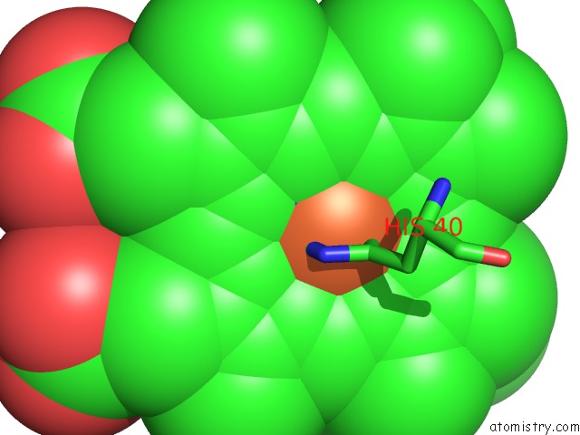
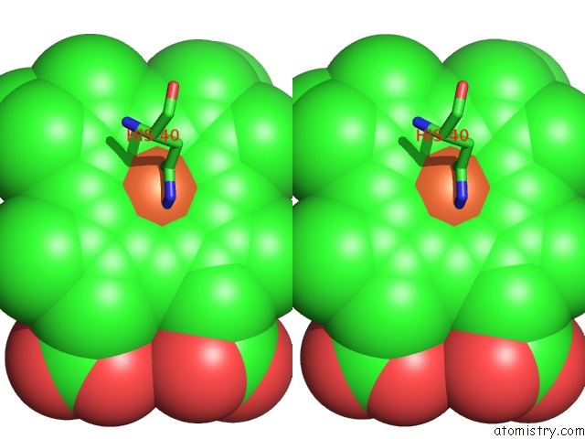
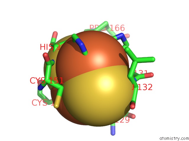
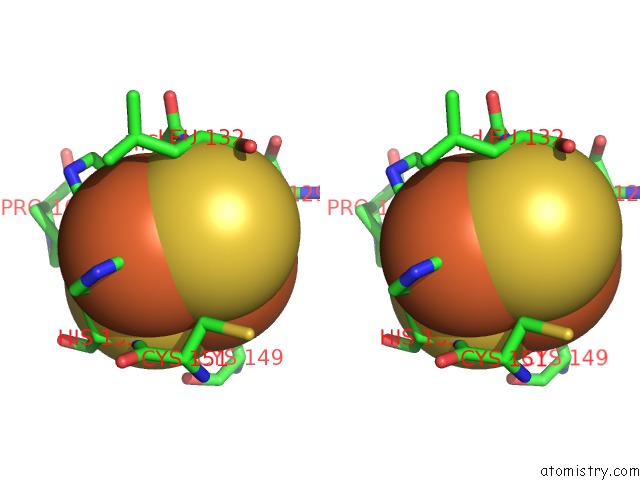
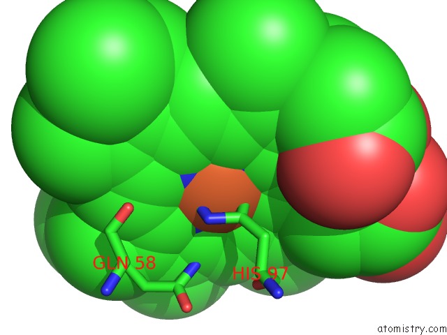
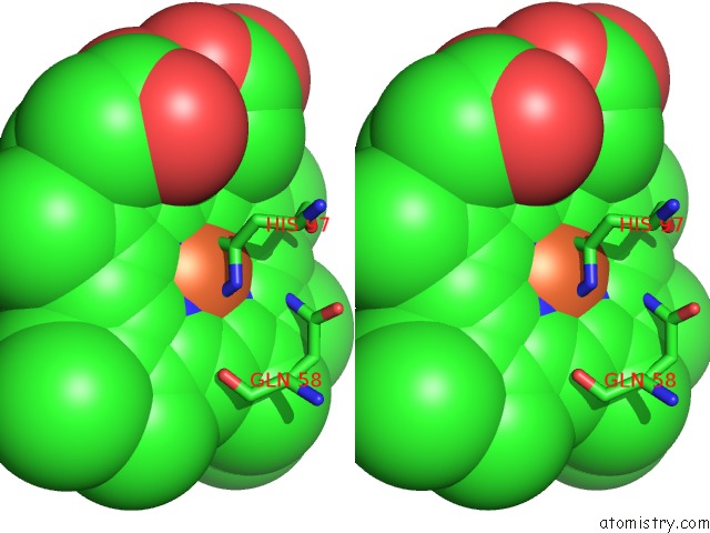
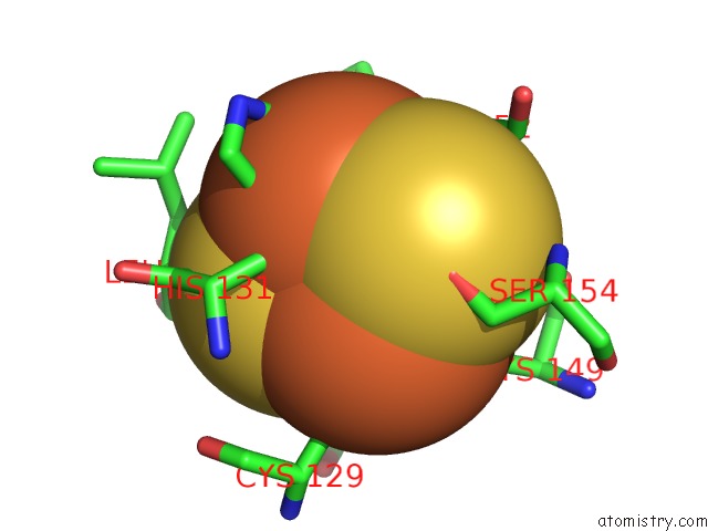
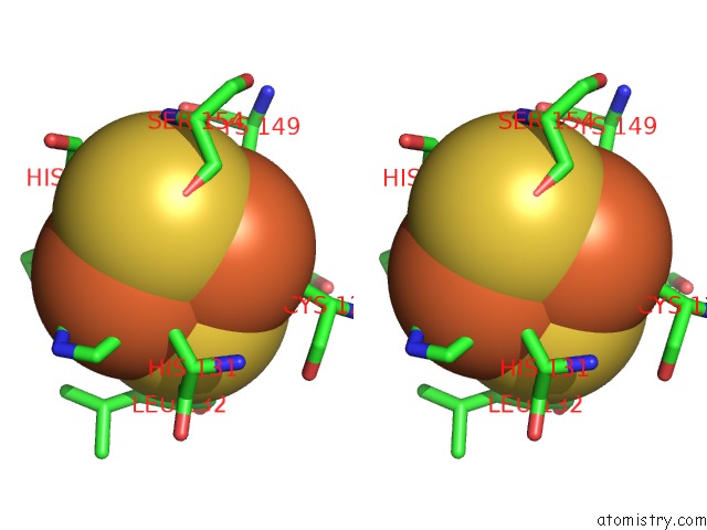
Iron binding site 1 out of 30 in 2qjy
Go back to
Iron binding site 1 out
of 30 in the Crystal Structure of Rhodobacter Sphaeroides Double Mutant with Stigmatellin and UQ2
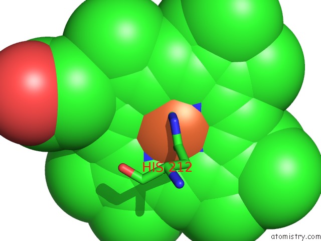
Mono view
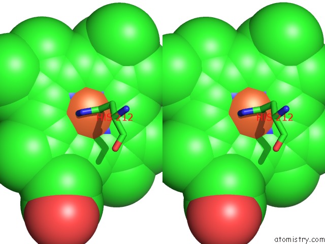
Stereo pair view
Mono view
Stereo pair view
A full contact list of Iron with other atoms in the Fe binding
site number 1 of Crystal Structure of Rhodobacter Sphaeroides Double Mutant with Stigmatellin and UQ2 within 5.0Å range:
|
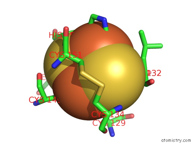
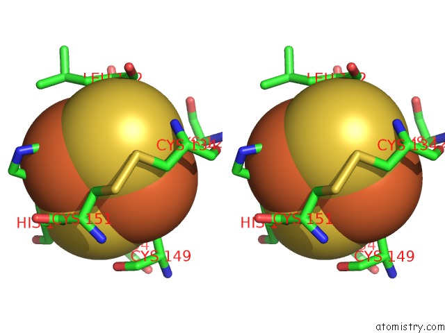
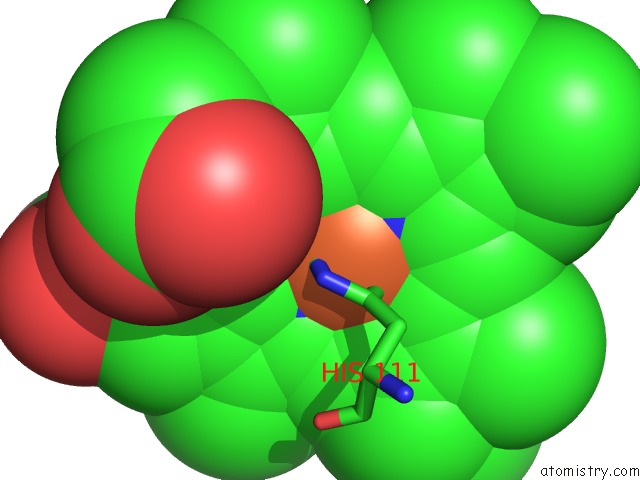
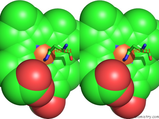
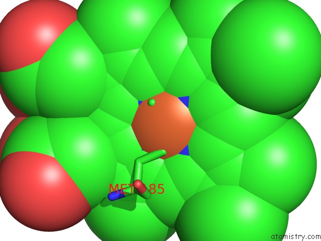
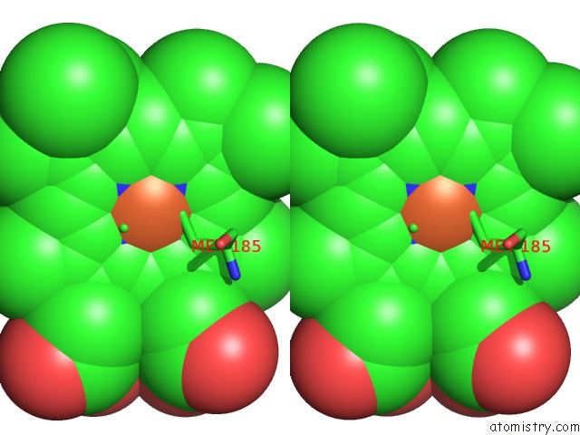
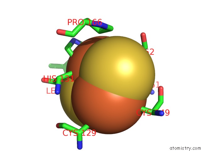
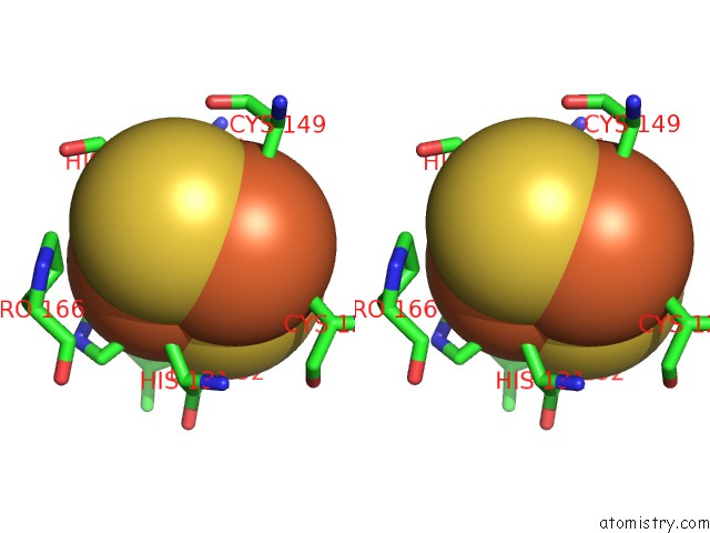
Iron binding site 2 out of 30 in 2qjy
Go back to
Iron binding site 2 out
of 30 in the Crystal Structure of Rhodobacter Sphaeroides Double Mutant with Stigmatellin and UQ2
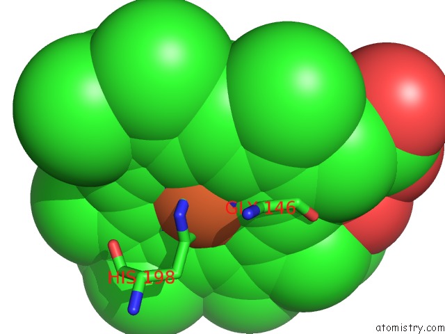
Mono view
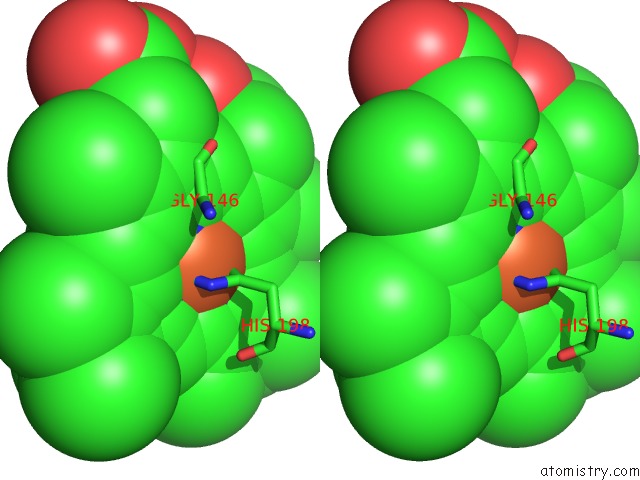
Stereo pair view
Mono view
Stereo pair view
A full contact list of Iron with other atoms in the Fe binding
site number 2 of Crystal Structure of Rhodobacter Sphaeroides Double Mutant with Stigmatellin and UQ2 within 5.0Å range:
|
Iron binding site 3 out of 30 in 2qjy
Go back to
Iron binding site 3 out
of 30 in the Crystal Structure of Rhodobacter Sphaeroides Double Mutant with Stigmatellin and UQ2
Mono view
Stereo pair view
Mono view
Stereo pair view
A full contact list of Iron with other atoms in the Fe binding
site number 3 of Crystal Structure of Rhodobacter Sphaeroides Double Mutant with Stigmatellin and UQ2 within 5.0Å range:
|
Iron binding site 4 out of 30 in 2qjy
Go back to
Iron binding site 4 out
of 30 in the Crystal Structure of Rhodobacter Sphaeroides Double Mutant with Stigmatellin and UQ2
Mono view
Stereo pair view
Mono view
Stereo pair view
A full contact list of Iron with other atoms in the Fe binding
site number 4 of Crystal Structure of Rhodobacter Sphaeroides Double Mutant with Stigmatellin and UQ2 within 5.0Å range:
|
Iron binding site 5 out of 30 in 2qjy
Go back to
Iron binding site 5 out
of 30 in the Crystal Structure of Rhodobacter Sphaeroides Double Mutant with Stigmatellin and UQ2
Mono view
Stereo pair view
Mono view
Stereo pair view
A full contact list of Iron with other atoms in the Fe binding
site number 5 of Crystal Structure of Rhodobacter Sphaeroides Double Mutant with Stigmatellin and UQ2 within 5.0Å range:
|
Iron binding site 6 out of 30 in 2qjy
Go back to
Iron binding site 6 out
of 30 in the Crystal Structure of Rhodobacter Sphaeroides Double Mutant with Stigmatellin and UQ2
Mono view
Stereo pair view
Mono view
Stereo pair view
A full contact list of Iron with other atoms in the Fe binding
site number 6 of Crystal Structure of Rhodobacter Sphaeroides Double Mutant with Stigmatellin and UQ2 within 5.0Å range:
|
Iron binding site 7 out of 30 in 2qjy
Go back to
Iron binding site 7 out
of 30 in the Crystal Structure of Rhodobacter Sphaeroides Double Mutant with Stigmatellin and UQ2
Mono view
Stereo pair view
Mono view
Stereo pair view
A full contact list of Iron with other atoms in the Fe binding
site number 7 of Crystal Structure of Rhodobacter Sphaeroides Double Mutant with Stigmatellin and UQ2 within 5.0Å range:
|
Iron binding site 8 out of 30 in 2qjy
Go back to
Iron binding site 8 out
of 30 in the Crystal Structure of Rhodobacter Sphaeroides Double Mutant with Stigmatellin and UQ2
Mono view
Stereo pair view
Mono view
Stereo pair view
A full contact list of Iron with other atoms in the Fe binding
site number 8 of Crystal Structure of Rhodobacter Sphaeroides Double Mutant with Stigmatellin and UQ2 within 5.0Å range:
|
Iron binding site 9 out of 30 in 2qjy
Go back to
Iron binding site 9 out
of 30 in the Crystal Structure of Rhodobacter Sphaeroides Double Mutant with Stigmatellin and UQ2
Mono view
Stereo pair view
Mono view
Stereo pair view
A full contact list of Iron with other atoms in the Fe binding
site number 9 of Crystal Structure of Rhodobacter Sphaeroides Double Mutant with Stigmatellin and UQ2 within 5.0Å range:
|
Iron binding site 10 out of 30 in 2qjy
Go back to
Iron binding site 10 out
of 30 in the Crystal Structure of Rhodobacter Sphaeroides Double Mutant with Stigmatellin and UQ2
Mono view
Stereo pair view
Mono view
Stereo pair view
A full contact list of Iron with other atoms in the Fe binding
site number 10 of Crystal Structure of Rhodobacter Sphaeroides Double Mutant with Stigmatellin and UQ2 within 5.0Å range:
|
Reference:
L.Esser,
M.Elberry,
F.Zhou,
C.A.Yu,
L.Yu,
D.Xia.
Inhibitor-Complexed Structures of the Cytochrome BC1 From the Photosynthetic Bacterium Rhodobacter Sphaeroides. J.Biol.Chem. V. 283 2846 2008.
ISSN: ISSN 0021-9258
PubMed: 18039651
DOI: 10.1074/JBC.M708608200
Page generated: Thu Jul 17 03:42:13 2025
ISSN: ISSN 0021-9258
PubMed: 18039651
DOI: 10.1074/JBC.M708608200
Last articles
Fe in 2YXOFe in 2YRS
Fe in 2YXC
Fe in 2YNM
Fe in 2YVJ
Fe in 2YP1
Fe in 2YU2
Fe in 2YU1
Fe in 2YQB
Fe in 2YOO