Iron »
PDB 2r1m-2rfb »
2rbw »
Iron in PDB 2rbw: Cytochrome C Peroxidase W191G in Complex with 1,2-Dimethyl-1H-Pyridin- 5-Amine
Enzymatic activity of Cytochrome C Peroxidase W191G in Complex with 1,2-Dimethyl-1H-Pyridin- 5-Amine
All present enzymatic activity of Cytochrome C Peroxidase W191G in Complex with 1,2-Dimethyl-1H-Pyridin- 5-Amine:
1.11.1.5;
1.11.1.5;
Protein crystallography data
The structure of Cytochrome C Peroxidase W191G in Complex with 1,2-Dimethyl-1H-Pyridin- 5-Amine, PDB code: 2rbw
was solved by
A.P.Graves,
S.E.Boyce,
B.K.Shoichet,
with X-Ray Crystallography technique. A brief refinement statistics is given in the table below:
| Resolution Low / High (Å) | 30.00 / 1.50 |
| Space group | P 21 21 21 |
| Cell size a, b, c (Å), α, β, γ (°) | 50.968, 75.044, 106.579, 90.00, 90.00, 90.00 |
| R / Rfree (%) | 14.4 / 16.7 |
Iron Binding Sites:
The binding sites of Iron atom in the Cytochrome C Peroxidase W191G in Complex with 1,2-Dimethyl-1H-Pyridin- 5-Amine
(pdb code 2rbw). This binding sites where shown within
5.0 Angstroms radius around Iron atom.
In total only one binding site of Iron was determined in the Cytochrome C Peroxidase W191G in Complex with 1,2-Dimethyl-1H-Pyridin- 5-Amine, PDB code: 2rbw:
In total only one binding site of Iron was determined in the Cytochrome C Peroxidase W191G in Complex with 1,2-Dimethyl-1H-Pyridin- 5-Amine, PDB code: 2rbw:
Iron binding site 1 out of 1 in 2rbw
Go back to
Iron binding site 1 out
of 1 in the Cytochrome C Peroxidase W191G in Complex with 1,2-Dimethyl-1H-Pyridin- 5-Amine
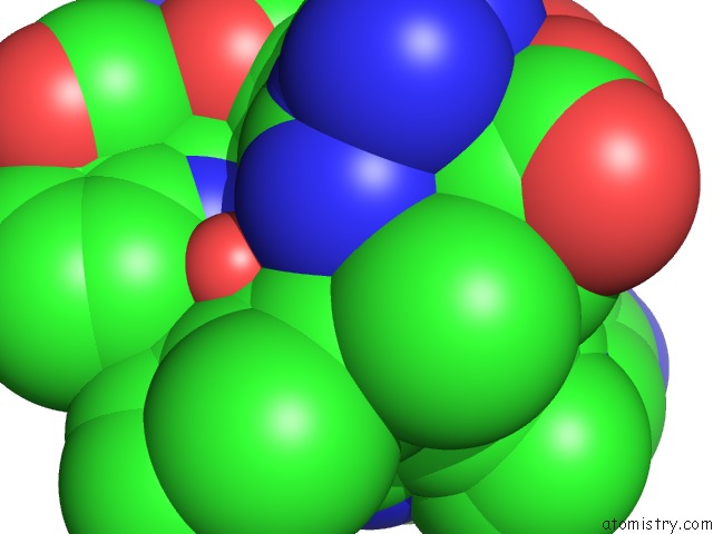
Mono view
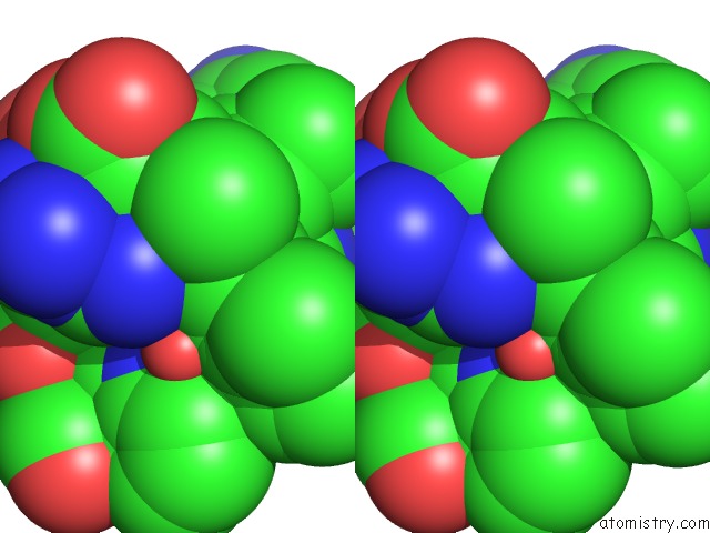
Stereo pair view
Mono view
Stereo pair view
A full contact list of Iron with other atoms in the Fe binding
site number 1 of Cytochrome C Peroxidase W191G in Complex with 1,2-Dimethyl-1H-Pyridin- 5-Amine within 5.0Å range:
|
Reference:
A.P.Graves,
D.M.Shivakumar,
S.E.Boyce,
M.P.Jacobson,
D.A.Case,
B.K.Shoichet.
Rescoring Docking Hit Lists For Model Cavity Sites: Predictions and Experimental Testing. J.Mol.Biol. V. 377 914 2008.
ISSN: ISSN 0022-2836
PubMed: 18280498
DOI: 10.1016/J.JMB.2008.01.049
Page generated: Thu Jul 17 03:56:01 2025
ISSN: ISSN 0022-2836
PubMed: 18280498
DOI: 10.1016/J.JMB.2008.01.049
Last articles
Fe in 2YXOFe in 2YRS
Fe in 2YXC
Fe in 2YNM
Fe in 2YVJ
Fe in 2YP1
Fe in 2YU2
Fe in 2YU1
Fe in 2YQB
Fe in 2YOO