Iron »
PDB 2r1m-2rfb »
2rc0 »
Iron in PDB 2rc0: Cytochrome C Peroxidase W191G in Complex with 2-Imino-4- Methylpiperdine
Enzymatic activity of Cytochrome C Peroxidase W191G in Complex with 2-Imino-4- Methylpiperdine
All present enzymatic activity of Cytochrome C Peroxidase W191G in Complex with 2-Imino-4- Methylpiperdine:
1.11.1.5;
1.11.1.5;
Protein crystallography data
The structure of Cytochrome C Peroxidase W191G in Complex with 2-Imino-4- Methylpiperdine, PDB code: 2rc0
was solved by
A.P.Graves,
S.E.Boyce,
B.K.Shoichet,
with X-Ray Crystallography technique. A brief refinement statistics is given in the table below:
| Resolution Low / High (Å) | 30.00 / 1.50 |
| Space group | P 21 21 21 |
| Cell size a, b, c (Å), α, β, γ (°) | 51.110, 75.961, 106.950, 90.00, 90.00, 90.00 |
| R / Rfree (%) | 14.3 / 16.9 |
Iron Binding Sites:
The binding sites of Iron atom in the Cytochrome C Peroxidase W191G in Complex with 2-Imino-4- Methylpiperdine
(pdb code 2rc0). This binding sites where shown within
5.0 Angstroms radius around Iron atom.
In total only one binding site of Iron was determined in the Cytochrome C Peroxidase W191G in Complex with 2-Imino-4- Methylpiperdine, PDB code: 2rc0:
In total only one binding site of Iron was determined in the Cytochrome C Peroxidase W191G in Complex with 2-Imino-4- Methylpiperdine, PDB code: 2rc0:
Iron binding site 1 out of 1 in 2rc0
Go back to
Iron binding site 1 out
of 1 in the Cytochrome C Peroxidase W191G in Complex with 2-Imino-4- Methylpiperdine
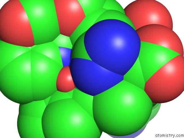
Mono view
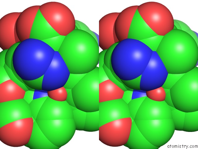
Stereo pair view
Mono view
Stereo pair view
A full contact list of Iron with other atoms in the Fe binding
site number 1 of Cytochrome C Peroxidase W191G in Complex with 2-Imino-4- Methylpiperdine within 5.0Å range:
|
Reference:
A.P.Graves,
D.M.Shivakumar,
S.E.Boyce,
M.P.Jacobson,
D.A.Case,
B.K.Shoichet.
Rescoring Docking Hit Lists For Model Cavity Sites: Predictions and Experimental Testing. J.Mol.Biol. V. 377 914 2008.
ISSN: ISSN 0022-2836
PubMed: 18280498
DOI: 10.1016/J.JMB.2008.01.049
Page generated: Sun Aug 4 02:08:34 2024
ISSN: ISSN 0022-2836
PubMed: 18280498
DOI: 10.1016/J.JMB.2008.01.049
Last articles
Zn in 9J0NZn in 9J0O
Zn in 9J0P
Zn in 9FJX
Zn in 9EKB
Zn in 9C0F
Zn in 9CAH
Zn in 9CH0
Zn in 9CH3
Zn in 9CH1