Iron »
PDB 2r1m-2rfb »
2rch »
Iron in PDB 2rch: Crystal Structure of Arabidopsis Thaliana Allene Oxide Synthase (Aos, Cytochrome P450 74A, CYP74A) Complexed with 13(S)-Hod at 1.85 A Resolution
Enzymatic activity of Crystal Structure of Arabidopsis Thaliana Allene Oxide Synthase (Aos, Cytochrome P450 74A, CYP74A) Complexed with 13(S)-Hod at 1.85 A Resolution
All present enzymatic activity of Crystal Structure of Arabidopsis Thaliana Allene Oxide Synthase (Aos, Cytochrome P450 74A, CYP74A) Complexed with 13(S)-Hod at 1.85 A Resolution:
4.2.1.92;
4.2.1.92;
Protein crystallography data
The structure of Crystal Structure of Arabidopsis Thaliana Allene Oxide Synthase (Aos, Cytochrome P450 74A, CYP74A) Complexed with 13(S)-Hod at 1.85 A Resolution, PDB code: 2rch
was solved by
D.S.Lee,
P.Nioche,
C.S.Raman,
with X-Ray Crystallography technique. A brief refinement statistics is given in the table below:
| Resolution Low / High (Å) | 88.39 / 1.85 |
| Space group | P 21 21 21 |
| Cell size a, b, c (Å), α, β, γ (°) | 63.552, 105.336, 162.503, 90.00, 90.00, 90.00 |
| R / Rfree (%) | 17.1 / 20.5 |
Iron Binding Sites:
The binding sites of Iron atom in the Crystal Structure of Arabidopsis Thaliana Allene Oxide Synthase (Aos, Cytochrome P450 74A, CYP74A) Complexed with 13(S)-Hod at 1.85 A Resolution
(pdb code 2rch). This binding sites where shown within
5.0 Angstroms radius around Iron atom.
In total 2 binding sites of Iron where determined in the Crystal Structure of Arabidopsis Thaliana Allene Oxide Synthase (Aos, Cytochrome P450 74A, CYP74A) Complexed with 13(S)-Hod at 1.85 A Resolution, PDB code: 2rch:
Jump to Iron binding site number: 1; 2;
In total 2 binding sites of Iron where determined in the Crystal Structure of Arabidopsis Thaliana Allene Oxide Synthase (Aos, Cytochrome P450 74A, CYP74A) Complexed with 13(S)-Hod at 1.85 A Resolution, PDB code: 2rch:
Jump to Iron binding site number: 1; 2;
Iron binding site 1 out of 2 in 2rch
Go back to
Iron binding site 1 out
of 2 in the Crystal Structure of Arabidopsis Thaliana Allene Oxide Synthase (Aos, Cytochrome P450 74A, CYP74A) Complexed with 13(S)-Hod at 1.85 A Resolution
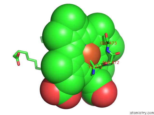
Mono view
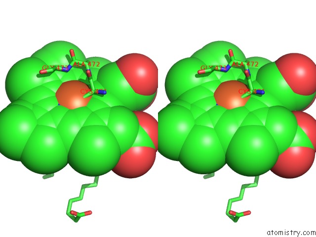
Stereo pair view
Mono view
Stereo pair view
A full contact list of Iron with other atoms in the Fe binding
site number 1 of Crystal Structure of Arabidopsis Thaliana Allene Oxide Synthase (Aos, Cytochrome P450 74A, CYP74A) Complexed with 13(S)-Hod at 1.85 A Resolution within 5.0Å range:
|
Iron binding site 2 out of 2 in 2rch
Go back to
Iron binding site 2 out
of 2 in the Crystal Structure of Arabidopsis Thaliana Allene Oxide Synthase (Aos, Cytochrome P450 74A, CYP74A) Complexed with 13(S)-Hod at 1.85 A Resolution
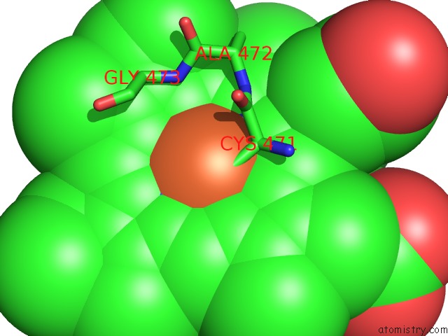
Mono view
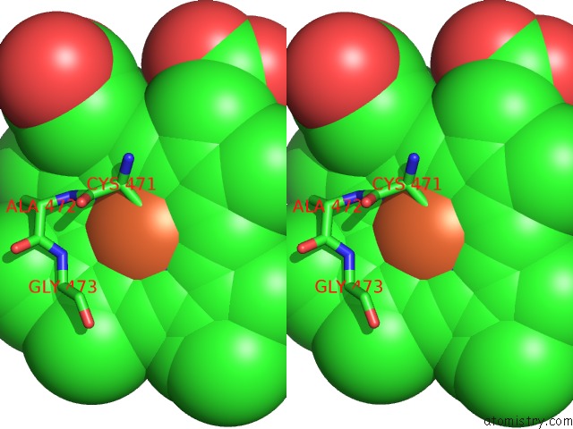
Stereo pair view
Mono view
Stereo pair view
A full contact list of Iron with other atoms in the Fe binding
site number 2 of Crystal Structure of Arabidopsis Thaliana Allene Oxide Synthase (Aos, Cytochrome P450 74A, CYP74A) Complexed with 13(S)-Hod at 1.85 A Resolution within 5.0Å range:
|
Reference:
D.S.Lee,
P.Nioche,
M.Hamberg,
C.S.Raman.
Structural Insights Into the Evolutionary Paths of Oxylipin Biosynthetic Enzymes. Nature V. 455 363 2008.
ISSN: ISSN 0028-0836
PubMed: 18716621
DOI: 10.1038/NATURE07307
Page generated: Sun Aug 4 02:09:00 2024
ISSN: ISSN 0028-0836
PubMed: 18716621
DOI: 10.1038/NATURE07307
Last articles
Zn in 9MJ5Zn in 9HNW
Zn in 9G0L
Zn in 9FNE
Zn in 9DZN
Zn in 9E0I
Zn in 9D32
Zn in 9DAK
Zn in 8ZXC
Zn in 8ZUF