Iron »
PDB 2r1m-2rfb »
2rdr »
Iron in PDB 2rdr: Crystal Structure of Ptlh with Fe/Oxalylglycine Bound
Protein crystallography data
The structure of Crystal Structure of Ptlh with Fe/Oxalylglycine Bound, PDB code: 2rdr
was solved by
Z.You,
S.Omura,
H.Ikeda,
D.E.Cane,
G.Jogl,
with X-Ray Crystallography technique. A brief refinement statistics is given in the table below:
| Resolution Low / High (Å) | 27.57 / 1.70 |
| Space group | P 1 21 1 |
| Cell size a, b, c (Å), α, β, γ (°) | 45.769, 70.564, 48.023, 90.00, 113.17, 90.00 |
| R / Rfree (%) | 17.4 / 21.7 |
Other elements in 2rdr:
The structure of Crystal Structure of Ptlh with Fe/Oxalylglycine Bound also contains other interesting chemical elements:
| Magnesium | (Mg) | 1 atom |
Iron Binding Sites:
The binding sites of Iron atom in the Crystal Structure of Ptlh with Fe/Oxalylglycine Bound
(pdb code 2rdr). This binding sites where shown within
5.0 Angstroms radius around Iron atom.
In total only one binding site of Iron was determined in the Crystal Structure of Ptlh with Fe/Oxalylglycine Bound, PDB code: 2rdr:
In total only one binding site of Iron was determined in the Crystal Structure of Ptlh with Fe/Oxalylglycine Bound, PDB code: 2rdr:
Iron binding site 1 out of 1 in 2rdr
Go back to
Iron binding site 1 out
of 1 in the Crystal Structure of Ptlh with Fe/Oxalylglycine Bound

Mono view
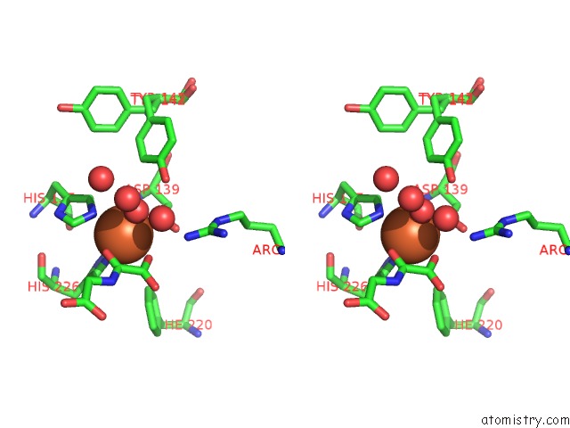
Stereo pair view
Mono view
Stereo pair view
A full contact list of Iron with other atoms in the Fe binding
site number 1 of Crystal Structure of Ptlh with Fe/Oxalylglycine Bound within 5.0Å range:
|
Reference:
Z.You,
S.Omura,
H.Ikeda,
D.E.Cane,
G.Jogl.
Crystal Structure of the Non-Heme Iron Dioxygenase Ptlh in Pentalenolactone Biosynthesis. J.Biol.Chem. V. 282 36552 2007.
ISSN: ISSN 0021-9258
PubMed: 17942405
DOI: 10.1074/JBC.M706358200
Page generated: Sun Aug 4 02:10:22 2024
ISSN: ISSN 0021-9258
PubMed: 17942405
DOI: 10.1074/JBC.M706358200
Last articles
Zn in 9MJ5Zn in 9HNW
Zn in 9G0L
Zn in 9FNE
Zn in 9DZN
Zn in 9E0I
Zn in 9D32
Zn in 9DAK
Zn in 8ZXC
Zn in 8ZUF