Iron »
PDB 2r1m-2rfb »
2rf7 »
Iron in PDB 2rf7: Crystal Structure of the Escherichia Coli Nrfa Mutant Q263E
Enzymatic activity of Crystal Structure of the Escherichia Coli Nrfa Mutant Q263E
All present enzymatic activity of Crystal Structure of the Escherichia Coli Nrfa Mutant Q263E:
1.7.2.2;
1.7.2.2;
Protein crystallography data
The structure of Crystal Structure of the Escherichia Coli Nrfa Mutant Q263E, PDB code: 2rf7
was solved by
T.A.Clarke,
D.J.Richardson,
A.M.Hemmings,
with X-Ray Crystallography technique. A brief refinement statistics is given in the table below:
| Resolution Low / High (Å) | 79.31 / 2.04 |
| Space group | P 21 21 21 |
| Cell size a, b, c (Å), α, β, γ (°) | 82.292, 91.201, 295.486, 90.00, 90.00, 90.00 |
| R / Rfree (%) | 17.1 / 22.6 |
Other elements in 2rf7:
The structure of Crystal Structure of the Escherichia Coli Nrfa Mutant Q263E also contains other interesting chemical elements:
| Calcium | (Ca) | 8 atoms |
Iron Binding Sites:
Pages:
>>> Page 1 <<< Page 2, Binding sites: 11 - 20;Binding sites:
The binding sites of Iron atom in the Crystal Structure of the Escherichia Coli Nrfa Mutant Q263E (pdb code 2rf7). This binding sites where shown within 5.0 Angstroms radius around Iron atom.In total 20 binding sites of Iron where determined in the Crystal Structure of the Escherichia Coli Nrfa Mutant Q263E, PDB code: 2rf7:
Jump to Iron binding site number: 1; 2; 3; 4; 5; 6; 7; 8; 9; 10;
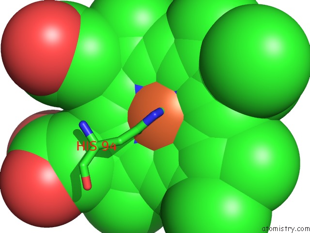
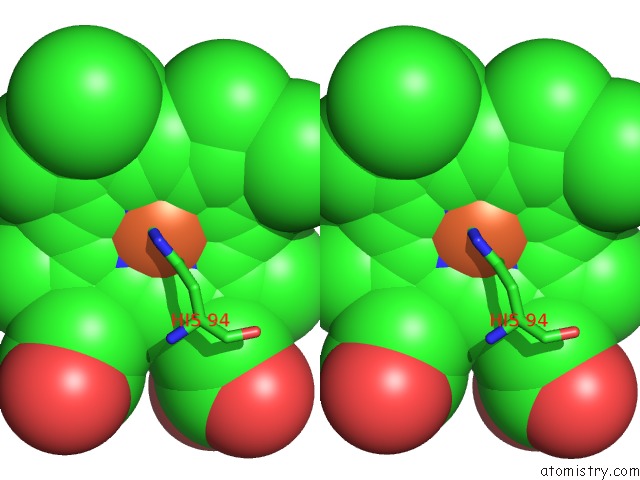
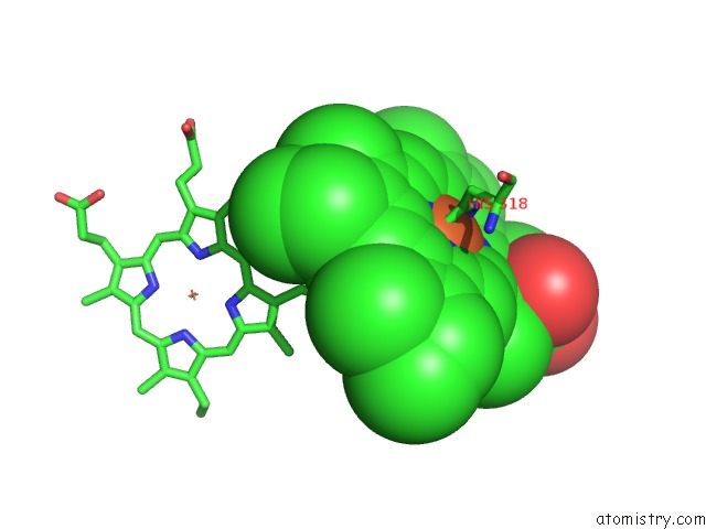
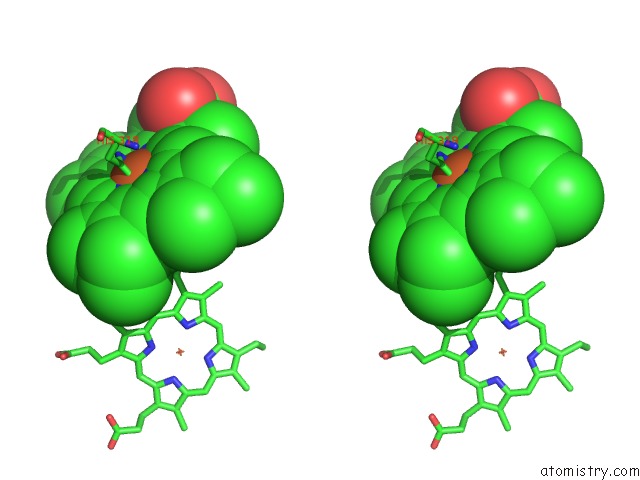
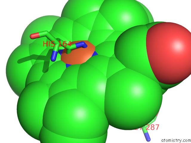
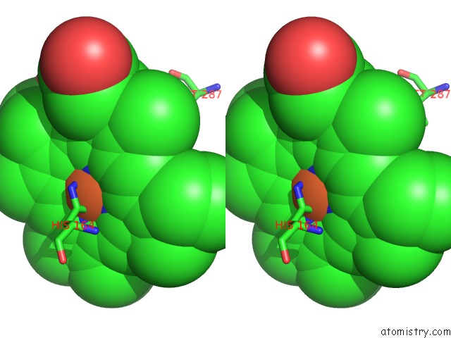
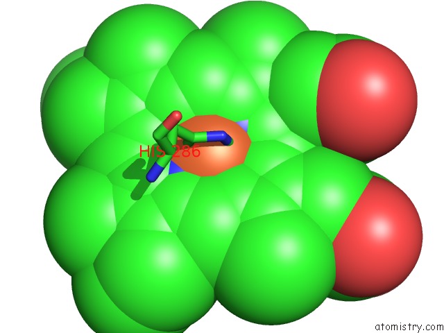
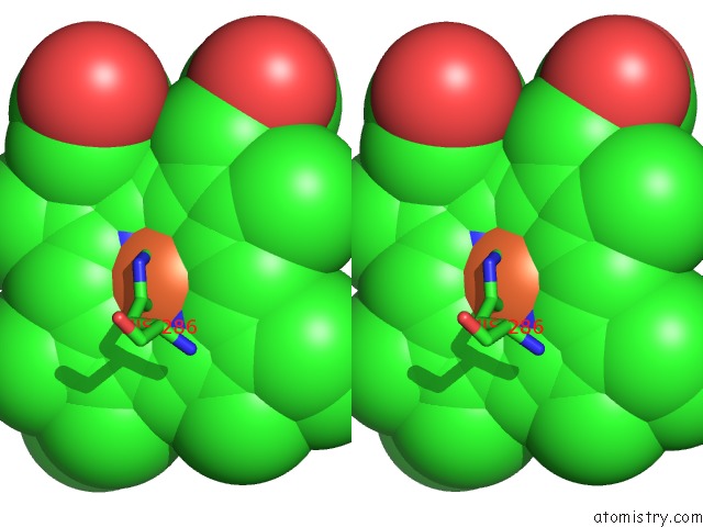
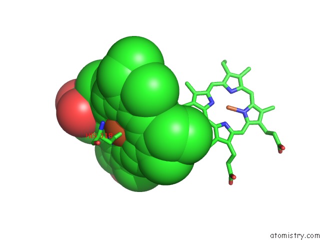
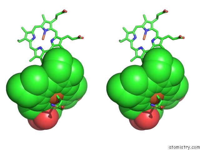
Iron binding site 1 out of 20 in 2rf7
Go back to
Iron binding site 1 out
of 20 in the Crystal Structure of the Escherichia Coli Nrfa Mutant Q263E
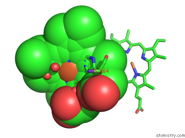
Mono view
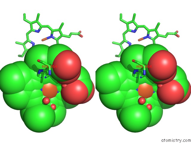
Stereo pair view
Mono view
Stereo pair view
A full contact list of Iron with other atoms in the Fe binding
site number 1 of Crystal Structure of the Escherichia Coli Nrfa Mutant Q263E within 5.0Å range:
|
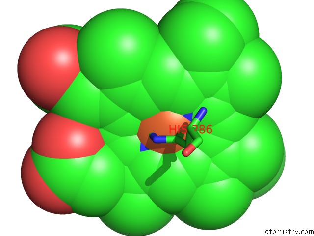
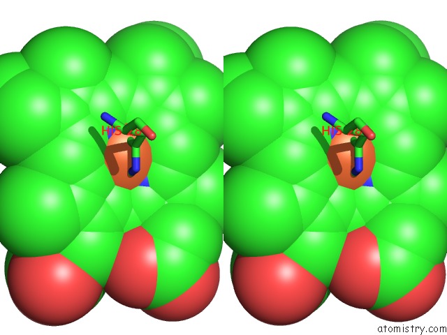
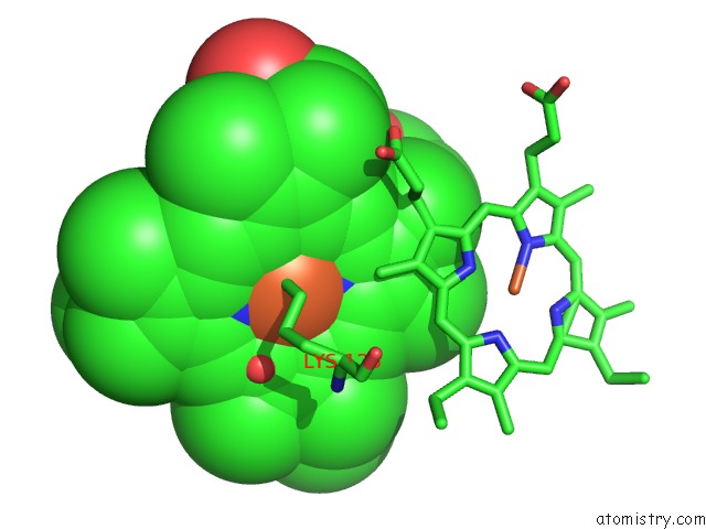
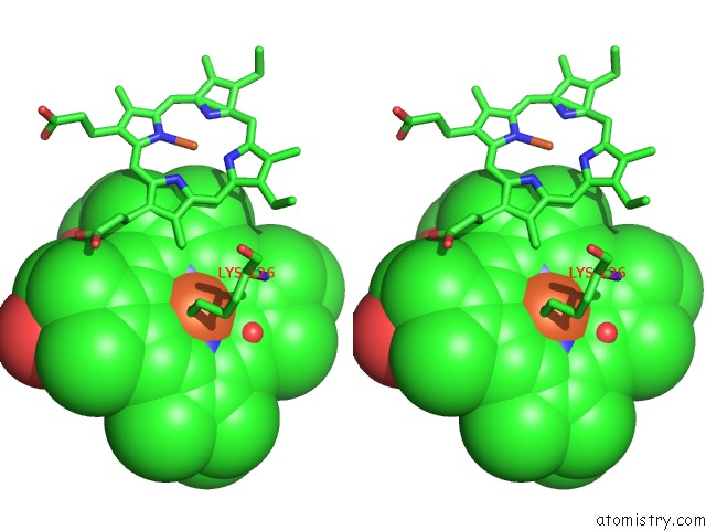
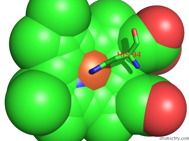
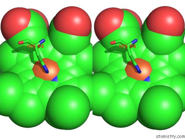
Iron binding site 2 out of 20 in 2rf7
Go back to
Iron binding site 2 out
of 20 in the Crystal Structure of the Escherichia Coli Nrfa Mutant Q263E
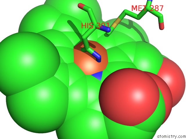
Mono view
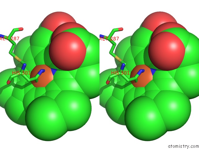
Stereo pair view
Mono view
Stereo pair view
A full contact list of Iron with other atoms in the Fe binding
site number 2 of Crystal Structure of the Escherichia Coli Nrfa Mutant Q263E within 5.0Å range:
|
Iron binding site 3 out of 20 in 2rf7
Go back to
Iron binding site 3 out
of 20 in the Crystal Structure of the Escherichia Coli Nrfa Mutant Q263E
Mono view
Stereo pair view
Mono view
Stereo pair view
A full contact list of Iron with other atoms in the Fe binding
site number 3 of Crystal Structure of the Escherichia Coli Nrfa Mutant Q263E within 5.0Å range:
|
Iron binding site 4 out of 20 in 2rf7
Go back to
Iron binding site 4 out
of 20 in the Crystal Structure of the Escherichia Coli Nrfa Mutant Q263E
Mono view
Stereo pair view
Mono view
Stereo pair view
A full contact list of Iron with other atoms in the Fe binding
site number 4 of Crystal Structure of the Escherichia Coli Nrfa Mutant Q263E within 5.0Å range:
|
Iron binding site 5 out of 20 in 2rf7
Go back to
Iron binding site 5 out
of 20 in the Crystal Structure of the Escherichia Coli Nrfa Mutant Q263E
Mono view
Stereo pair view
Mono view
Stereo pair view
A full contact list of Iron with other atoms in the Fe binding
site number 5 of Crystal Structure of the Escherichia Coli Nrfa Mutant Q263E within 5.0Å range:
|
Iron binding site 6 out of 20 in 2rf7
Go back to
Iron binding site 6 out
of 20 in the Crystal Structure of the Escherichia Coli Nrfa Mutant Q263E
Mono view
Stereo pair view
Mono view
Stereo pair view
A full contact list of Iron with other atoms in the Fe binding
site number 6 of Crystal Structure of the Escherichia Coli Nrfa Mutant Q263E within 5.0Å range:
|
Iron binding site 7 out of 20 in 2rf7
Go back to
Iron binding site 7 out
of 20 in the Crystal Structure of the Escherichia Coli Nrfa Mutant Q263E
Mono view
Stereo pair view
Mono view
Stereo pair view
A full contact list of Iron with other atoms in the Fe binding
site number 7 of Crystal Structure of the Escherichia Coli Nrfa Mutant Q263E within 5.0Å range:
|
Iron binding site 8 out of 20 in 2rf7
Go back to
Iron binding site 8 out
of 20 in the Crystal Structure of the Escherichia Coli Nrfa Mutant Q263E
Mono view
Stereo pair view
Mono view
Stereo pair view
A full contact list of Iron with other atoms in the Fe binding
site number 8 of Crystal Structure of the Escherichia Coli Nrfa Mutant Q263E within 5.0Å range:
|
Iron binding site 9 out of 20 in 2rf7
Go back to
Iron binding site 9 out
of 20 in the Crystal Structure of the Escherichia Coli Nrfa Mutant Q263E
Mono view
Stereo pair view
Mono view
Stereo pair view
A full contact list of Iron with other atoms in the Fe binding
site number 9 of Crystal Structure of the Escherichia Coli Nrfa Mutant Q263E within 5.0Å range:
|
Iron binding site 10 out of 20 in 2rf7
Go back to
Iron binding site 10 out
of 20 in the Crystal Structure of the Escherichia Coli Nrfa Mutant Q263E
Mono view
Stereo pair view
Mono view
Stereo pair view
A full contact list of Iron with other atoms in the Fe binding
site number 10 of Crystal Structure of the Escherichia Coli Nrfa Mutant Q263E within 5.0Å range:
|
Reference:
T.A.Clarke,
G.L.Kemp,
J.H.Wonderen,
R.M.Doyle,
J.A.Cole,
N.Tovell,
M.R.Cheesman,
J.N.Butt,
D.J.Richardson,
A.M.Hemmings.
Role of A Conserved Glutamine Residue in Tuning the Catalytic Activity of Escherichia Coli Cytochrome C Nitrite Reductase. Biochemistry V. 47 3789 2008.
ISSN: ISSN 0006-2960
PubMed: 18311941
DOI: 10.1021/BI702175W
Page generated: Thu Jul 17 03:57:57 2025
ISSN: ISSN 0006-2960
PubMed: 18311941
DOI: 10.1021/BI702175W
Last articles
Fe in 2YXOFe in 2YRS
Fe in 2YXC
Fe in 2YNM
Fe in 2YVJ
Fe in 2YP1
Fe in 2YU2
Fe in 2YU1
Fe in 2YQB
Fe in 2YOO