Iron »
PDB 2r1m-2rfb »
2rfb »
Iron in PDB 2rfb: Crystal Structure of A Cytochrome P450 From the Thermoacidophilic Archaeon Picrophilus Torridus
Enzymatic activity of Crystal Structure of A Cytochrome P450 From the Thermoacidophilic Archaeon Picrophilus Torridus
All present enzymatic activity of Crystal Structure of A Cytochrome P450 From the Thermoacidophilic Archaeon Picrophilus Torridus:
1.14.14.1;
1.14.14.1;
Protein crystallography data
The structure of Crystal Structure of A Cytochrome P450 From the Thermoacidophilic Archaeon Picrophilus Torridus, PDB code: 2rfb
was solved by
W.W.Ho,
H.Li,
T.L.Poulos,
C.R.Nishida,
P.R.Ortiz De Montellano,
with X-Ray Crystallography technique. A brief refinement statistics is given in the table below:
| Resolution Low / High (Å) | 48.51 / 2.50 |
| Space group | P 1 21 1 |
| Cell size a, b, c (Å), α, β, γ (°) | 48.790, 168.460, 88.230, 90.00, 96.25, 90.00 |
| R / Rfree (%) | 21.1 / 26.8 |
Iron Binding Sites:
The binding sites of Iron atom in the Crystal Structure of A Cytochrome P450 From the Thermoacidophilic Archaeon Picrophilus Torridus
(pdb code 2rfb). This binding sites where shown within
5.0 Angstroms radius around Iron atom.
In total 3 binding sites of Iron where determined in the Crystal Structure of A Cytochrome P450 From the Thermoacidophilic Archaeon Picrophilus Torridus, PDB code: 2rfb:
Jump to Iron binding site number: 1; 2; 3;
In total 3 binding sites of Iron where determined in the Crystal Structure of A Cytochrome P450 From the Thermoacidophilic Archaeon Picrophilus Torridus, PDB code: 2rfb:
Jump to Iron binding site number: 1; 2; 3;
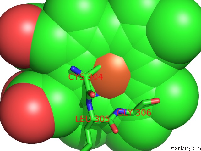
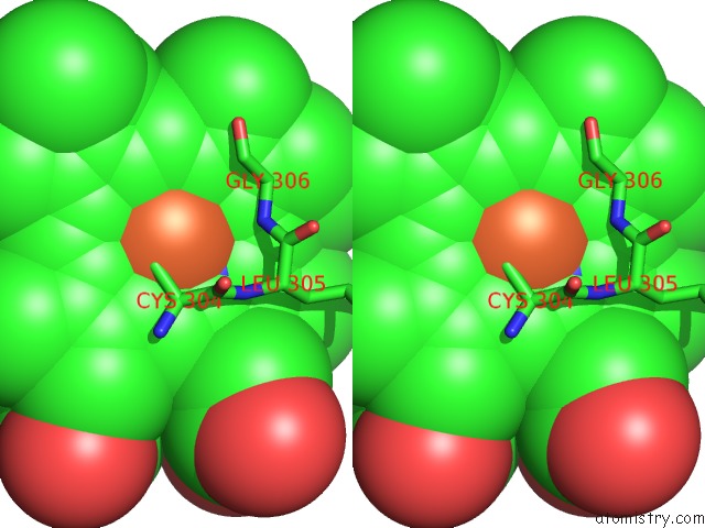
Iron binding site 1 out of 3 in 2rfb
Go back to
Iron binding site 1 out
of 3 in the Crystal Structure of A Cytochrome P450 From the Thermoacidophilic Archaeon Picrophilus Torridus
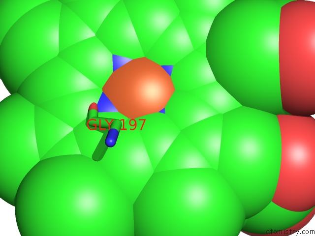
Mono view
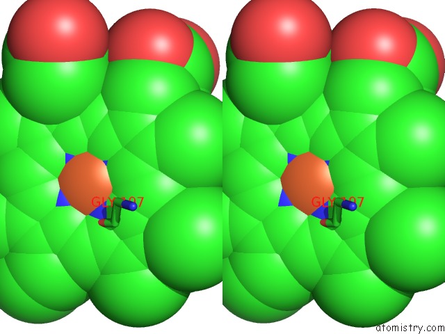
Stereo pair view
Mono view
Stereo pair view
A full contact list of Iron with other atoms in the Fe binding
site number 1 of Crystal Structure of A Cytochrome P450 From the Thermoacidophilic Archaeon Picrophilus Torridus within 5.0Å range:
|
Iron binding site 2 out of 3 in 2rfb
Go back to
Iron binding site 2 out
of 3 in the Crystal Structure of A Cytochrome P450 From the Thermoacidophilic Archaeon Picrophilus Torridus
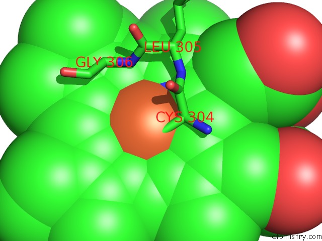
Mono view
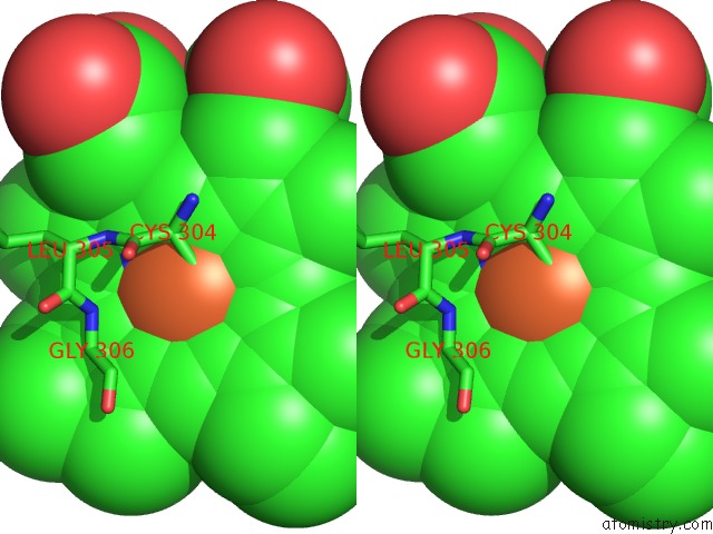
Stereo pair view
Mono view
Stereo pair view
A full contact list of Iron with other atoms in the Fe binding
site number 2 of Crystal Structure of A Cytochrome P450 From the Thermoacidophilic Archaeon Picrophilus Torridus within 5.0Å range:
|
Iron binding site 3 out of 3 in 2rfb
Go back to
Iron binding site 3 out
of 3 in the Crystal Structure of A Cytochrome P450 From the Thermoacidophilic Archaeon Picrophilus Torridus
Mono view
Stereo pair view
Mono view
Stereo pair view
A full contact list of Iron with other atoms in the Fe binding
site number 3 of Crystal Structure of A Cytochrome P450 From the Thermoacidophilic Archaeon Picrophilus Torridus within 5.0Å range:
|
Reference:
W.W.Ho,
H.Li,
C.R.Nishida,
P.R.Ortiz De Montellano,
T.L.Poulos.
Crystal Structure and Properties of CYP231A2 From the Thermoacidophilic Archaeon Picrophilus Torridus. Biochemistry V. 47 2071 2008.
ISSN: ISSN 0006-2960
PubMed: 18197710
DOI: 10.1021/BI702240K
Page generated: Sun Aug 4 02:11:07 2024
ISSN: ISSN 0006-2960
PubMed: 18197710
DOI: 10.1021/BI702240K
Last articles
Zn in 9J0NZn in 9J0O
Zn in 9J0P
Zn in 9FJX
Zn in 9EKB
Zn in 9C0F
Zn in 9CAH
Zn in 9CH0
Zn in 9CH3
Zn in 9CH1