Iron »
PDB 2rfc-2v1i »
2spm »
Iron in PDB 2spm: A Novel Site-Directed Mutant of Myoglobin with An Unusually High O2 Affinity and Low Autooxidation Rate
Protein crystallography data
The structure of A Novel Site-Directed Mutant of Myoglobin with An Unusually High O2 Affinity and Low Autooxidation Rate, PDB code: 2spm
was solved by
M.L.Quillin,
R.M.Arduini,
G.N.Phillips Jr.,
with X-Ray Crystallography technique. A brief refinement statistics is given in the table below:
| Resolution Low / High (Å) | 5.00 / 1.70 |
| Space group | P 6 |
| Cell size a, b, c (Å), α, β, γ (°) | 91.200, 91.200, 45.870, 90.00, 90.00, 120.00 |
| R / Rfree (%) | 16.7 / n/a |
Iron Binding Sites:
The binding sites of Iron atom in the A Novel Site-Directed Mutant of Myoglobin with An Unusually High O2 Affinity and Low Autooxidation Rate
(pdb code 2spm). This binding sites where shown within
5.0 Angstroms radius around Iron atom.
In total only one binding site of Iron was determined in the A Novel Site-Directed Mutant of Myoglobin with An Unusually High O2 Affinity and Low Autooxidation Rate, PDB code: 2spm:
In total only one binding site of Iron was determined in the A Novel Site-Directed Mutant of Myoglobin with An Unusually High O2 Affinity and Low Autooxidation Rate, PDB code: 2spm:
Iron binding site 1 out of 1 in 2spm
Go back to
Iron binding site 1 out
of 1 in the A Novel Site-Directed Mutant of Myoglobin with An Unusually High O2 Affinity and Low Autooxidation Rate
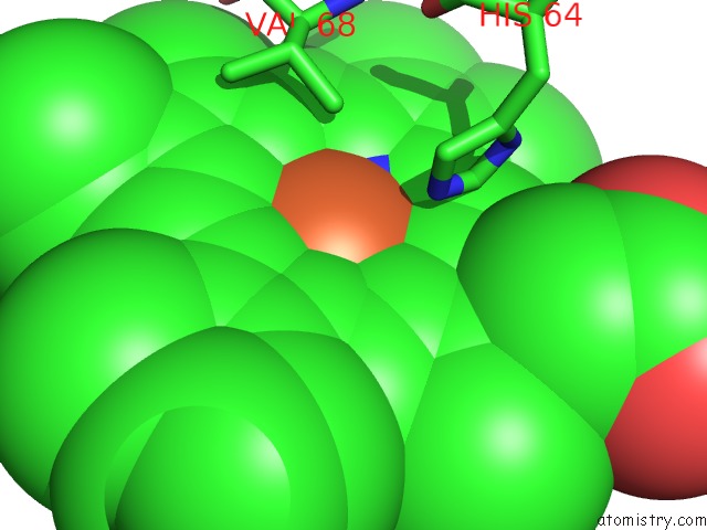
Mono view
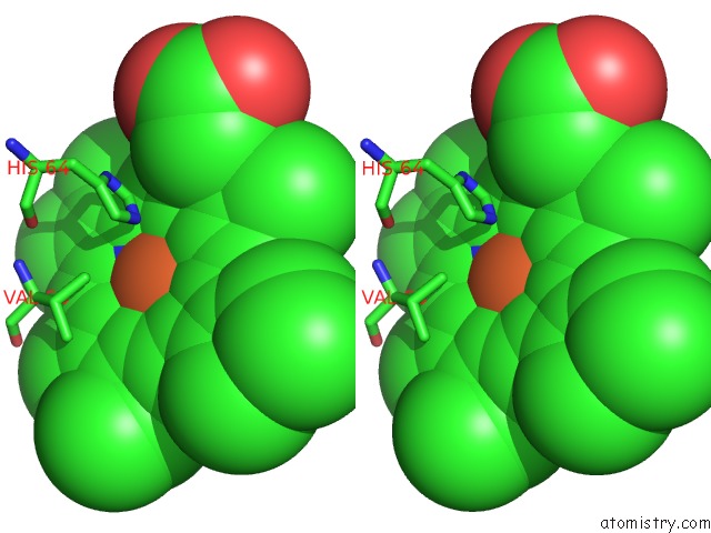
Stereo pair view
Mono view
Stereo pair view
A full contact list of Iron with other atoms in the Fe binding
site number 1 of A Novel Site-Directed Mutant of Myoglobin with An Unusually High O2 Affinity and Low Autooxidation Rate within 5.0Å range:
|
Reference:
T.E.Carver,
R.E.Brantley Jr.,
E.W.Singleton,
R.M.Arduini,
M.L.Quillin,
G.N.Phillips Jr.,
J.S.Olson.
A Novel Site-Directed Mutant of Myoglobin with An Unusually High O2 Affinity and Low Autooxidation Rate. J.Biol.Chem. V. 267 14443 1992.
ISSN: ISSN 0021-9258
PubMed: 1629229
Page generated: Thu Jul 17 04:02:11 2025
ISSN: ISSN 0021-9258
PubMed: 1629229
Last articles
Fe in 2YXOFe in 2YRS
Fe in 2YXC
Fe in 2YNM
Fe in 2YVJ
Fe in 2YP1
Fe in 2YU2
Fe in 2YU1
Fe in 2YQB
Fe in 2YOO