Atomistry » Iron » PDB 2rfc-2v1i » 2tmd
Atomistry »
  Iron »
    PDB 2rfc-2v1i »
      2tmd »

Iron in PDB 2tmd: Correlation of X-Ray Deduced and Experimental Amino Acid Sequences of Trimethylamine Dehydrogenase

Enzymatic activity of Correlation of X-Ray Deduced and Experimental Amino Acid Sequences of Trimethylamine Dehydrogenase

All present enzymatic activity of Correlation of X-Ray Deduced and Experimental Amino Acid Sequences of Trimethylamine Dehydrogenase:
1.5.99.7;

Protein crystallography data

The structure of Correlation of X-Ray Deduced and Experimental Amino Acid Sequences of Trimethylamine Dehydrogenase, PDB code: 2tmd was solved by F.S.Mathews, L.W.Lim, S.White, with X-Ray Crystallography technique. A brief refinement statistics is given in the table below:

Resolution Low / High (Å) 10.00 / 2.40
Space group P 1 21 1
Cell size a, b, c (Å), α, β, γ (°) 147.750, 71.950, 83.820, 90.00, 97.69, 90.00
R / Rfree (%) 15.4 / n/a

Iron Binding Sites:

The binding sites of Iron atom in the Correlation of X-Ray Deduced and Experimental Amino Acid Sequences of Trimethylamine Dehydrogenase (pdb code 2tmd). This binding sites where shown within 5.0 Angstroms radius around Iron atom.
In total 8 binding sites of Iron where determined in the Correlation of X-Ray Deduced and Experimental Amino Acid Sequences of Trimethylamine Dehydrogenase, PDB code: 2tmd:
Jump to Iron binding site number: 1; 2; 3; 4; 5; 6; 7; 8;

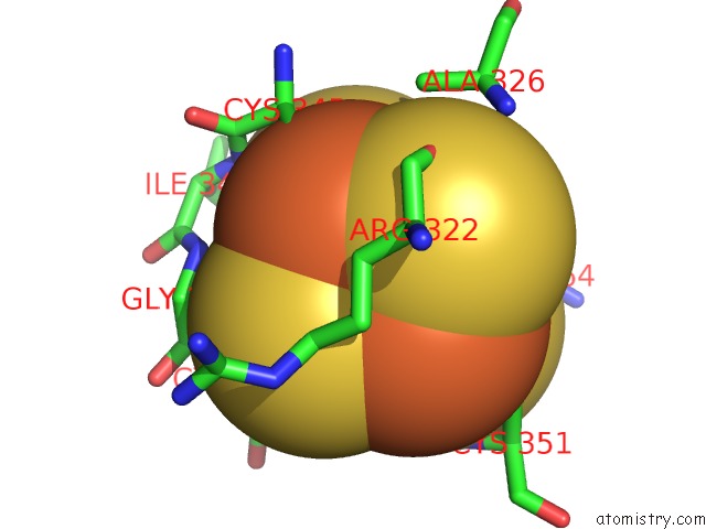
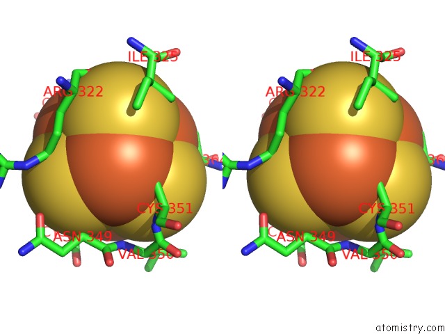
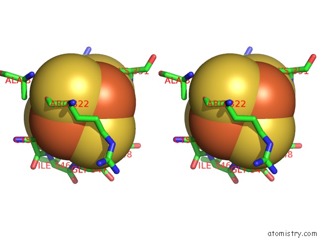
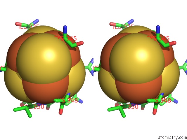
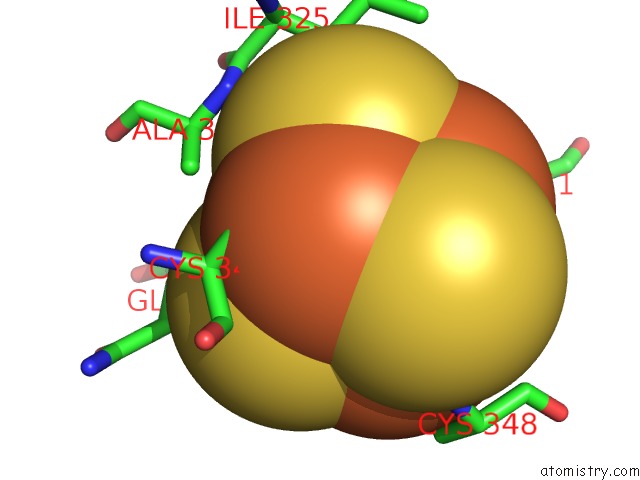
Iron binding site 1 out of 8 in 2tmd

Go back to Iron Binding Sites List in 2tmd
Iron binding site 1 out of 8 in the Correlation of X-Ray Deduced and Experimental Amino Acid Sequences of Trimethylamine Dehydrogenase


Mono view


Stereo pair view

A full contact list of Iron with other atoms in the Fe binding site number 1 of Correlation of X-Ray Deduced and Experimental Amino Acid Sequences of Trimethylamine Dehydrogenase within 5.0Å range:
probe atom residue distance (Å) B Occ
A:Fe801

b:12.1
occ:1.00
FE1 A:SF4801 0.0 12.1 1.0
SG A:CYS345 2.3 7.9 1.0
S2 A:SF4801 2.3 12.7 1.0
S4 A:SF4801 2.3 14.2 1.0
S3 A:SF4801 2.3 13.5 1.0
FE4 A:SF4801 2.7 14.9 1.0
FE3 A:SF4801 2.7 13.4 1.0
FE2 A:SF4801 2.8 18.3 1.0
CB A:CYS345 3.4 13.1 1.0
CA A:CYS345 3.8 11.6 1.0
S1 A:SF4801 3.9 13.5 1.0
N A:ILE346 4.0 10.1 1.0
N A:GLY347 4.1 10.8 1.0
CB A:ARG322 4.1 3.6 1.0
C A:CYS345 4.3 10.2 1.0
CD A:ARG322 4.4 7.7 1.0
O A:ARG322 4.4 10.9 1.0
CB A:ALA326 4.5 7.5 1.0
CA A:GLY347 4.7 8.0 1.0
SG A:CYS364 4.7 10.9 1.0
CG A:ARG322 4.7 6.1 1.0
C A:ARG322 4.8 7.1 1.0
N A:CYS348 4.8 6.8 1.0
SG A:CYS351 4.9 8.2 1.0
CA A:ARG322 4.9 7.5 1.0
C A:ILE346 5.0 11.3 1.0

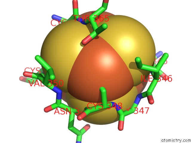
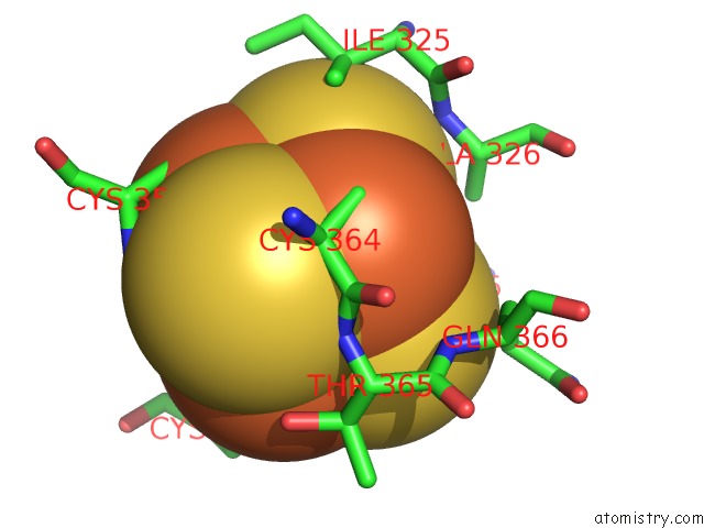
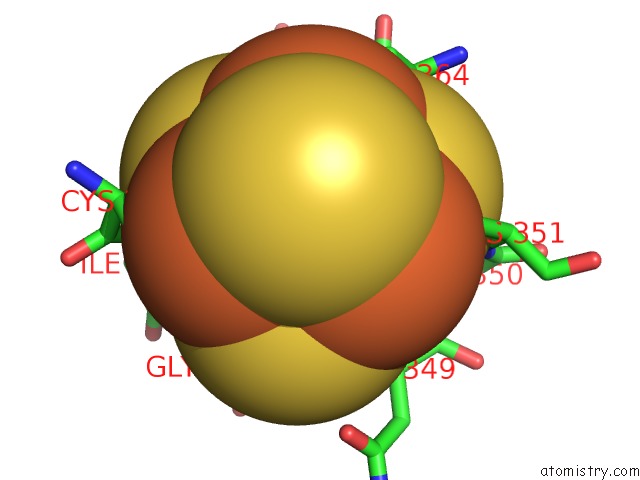
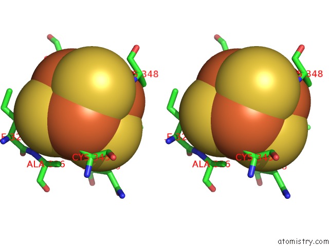
Iron binding site 2 out of 8 in 2tmd

Go back to Iron Binding Sites List in 2tmd
Iron binding site 2 out of 8 in the Correlation of X-Ray Deduced and Experimental Amino Acid Sequences of Trimethylamine Dehydrogenase


Mono view


Stereo pair view

A full contact list of Iron with other atoms in the Fe binding site number 2 of Correlation of X-Ray Deduced and Experimental Amino Acid Sequences of Trimethylamine Dehydrogenase within 5.0Å range:
probe atom residue distance (Å) B Occ
A:Fe801

b:18.3
occ:1.00
FE2 A:SF4801 0.0 18.3 1.0
S1 A:SF4801 2.3 13.5 1.0
S3 A:SF4801 2.3 13.5 1.0
S4 A:SF4801 2.3 14.2 1.0
SG A:CYS348 2.3 11.0 1.0
FE4 A:SF4801 2.8 14.9 1.0
FE3 A:SF4801 2.8 13.4 1.0
FE1 A:SF4801 2.8 12.1 1.0
N A:CYS348 3.7 6.8 1.0
CB A:CYS348 3.7 6.6 1.0
N A:ASN349 3.8 8.3 1.0
OG1 A:THR365 4.0 11.8 1.0
S2 A:SF4801 4.0 12.7 1.0
CA A:CYS348 4.1 8.0 1.0
CB A:THR365 4.2 11.3 1.0
CG1 A:ILE346 4.2 9.4 1.0
C A:CYS348 4.3 9.8 1.0
N A:VAL350 4.3 7.5 1.0
N A:THR365 4.4 11.5 1.0
N A:GLY347 4.6 10.8 1.0
CA A:ASN349 4.7 7.0 1.0
SG A:CYS345 4.7 7.9 1.0
SG A:CYS351 4.8 8.2 1.0
C A:GLY347 4.8 10.1 1.0
SG A:CYS364 4.9 10.9 1.0
N A:ILE346 4.9 10.1 1.0
N A:CYS351 4.9 5.9 1.0
CA A:THR365 5.0 9.8 1.0
CD1 A:ILE346 5.0 9.1 1.0
CB A:VAL350 5.0 4.5 1.0
C A:ASN349 5.0 6.5 1.0

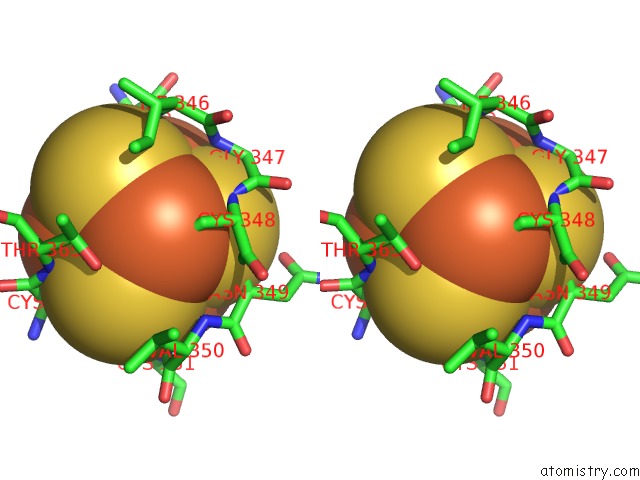
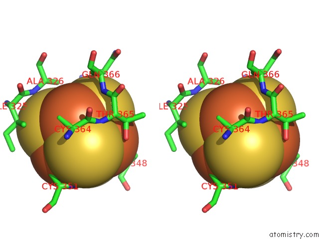
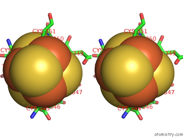
Iron binding site 3 out of 8 in 2tmd

Go back to Iron Binding Sites List in 2tmd
Iron binding site 3 out of 8 in the Correlation of X-Ray Deduced and Experimental Amino Acid Sequences of Trimethylamine Dehydrogenase


Mono view


Stereo pair view

A full contact list of Iron with other atoms in the Fe binding site number 3 of Correlation of X-Ray Deduced and Experimental Amino Acid Sequences of Trimethylamine Dehydrogenase within 5.0Å range:
probe atom residue distance (Å) B Occ
A:Fe801

b:13.4
occ:1.00
FE3 A:SF4801 0.0 13.4 1.0
S4 A:SF4801 2.3 14.2 1.0
S1 A:SF4801 2.3 13.5 1.0
S2 A:SF4801 2.3 12.7 1.0
SG A:CYS351 2.3 8.2 1.0
FE4 A:SF4801 2.6 14.9 1.0
FE1 A:SF4801 2.7 12.1 1.0
FE2 A:SF4801 2.8 18.3 1.0
CB A:CYS351 3.4 2.3 1.0
S3 A:SF4801 3.9 13.5 1.0
N A:CYS351 4.0 5.9 1.0
CD A:ARG322 4.2 7.7 1.0
CA A:CYS351 4.3 8.4 1.0
CG A:ARG322 4.4 6.1 1.0
N A:ASN349 4.5 8.3 1.0
CB A:ARG322 4.5 3.6 1.0
CA A:ASN349 4.6 7.0 1.0
SG A:CYS345 4.6 7.9 1.0
SG A:CYS364 4.7 10.9 1.0
N A:VAL350 4.7 7.5 1.0
SG A:CYS348 4.8 11.0 1.0
C A:ASN349 4.9 6.5 1.0
CG2 A:ILE325 5.0 4.7 1.0

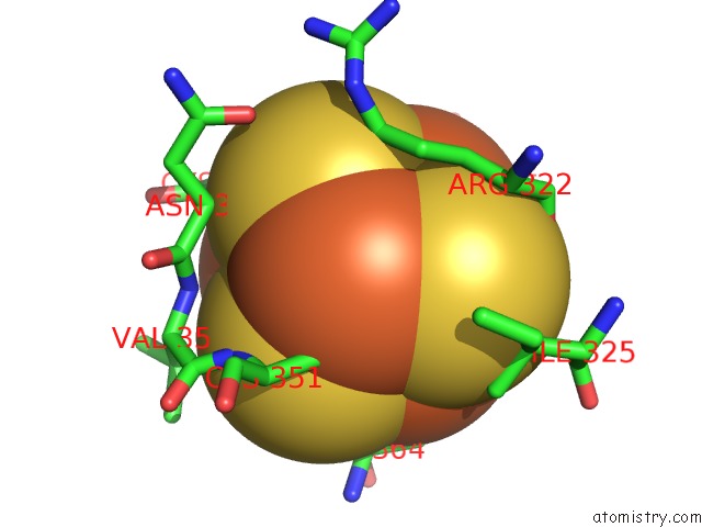
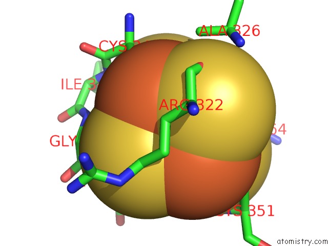
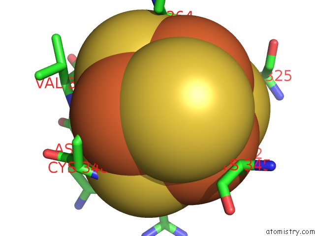
Iron binding site 4 out of 8 in 2tmd

Go back to Iron Binding Sites List in 2tmd
Iron binding site 4 out of 8 in the Correlation of X-Ray Deduced and Experimental Amino Acid Sequences of Trimethylamine Dehydrogenase


Mono view


Stereo pair view

A full contact list of Iron with other atoms in the Fe binding site number 4 of Correlation of X-Ray Deduced and Experimental Amino Acid Sequences of Trimethylamine Dehydrogenase within 5.0Å range:
probe atom residue distance (Å) B Occ
A:Fe801

b:14.9
occ:1.00
FE4 A:SF4801 0.0 14.9 1.0
S2 A:SF4801 2.3 12.7 1.0
S1 A:SF4801 2.3 13.5 1.0
SG A:CYS364 2.3 10.9 1.0
S3 A:SF4801 2.3 13.5 1.0
FE3 A:SF4801 2.6 13.4 1.0
FE1 A:SF4801 2.7 12.1 1.0
FE2 A:SF4801 2.8 18.3 1.0
CB A:CYS364 3.4 10.3 1.0
CA A:CYS364 3.7 12.8 1.0
N A:THR365 3.8 11.5 1.0
S4 A:SF4801 3.9 14.2 1.0
C A:CYS364 4.1 11.4 1.0
N A:GLN366 4.3 8.8 1.0
CB A:ALA326 4.5 7.5 1.0
CG2 A:ILE325 4.6 4.7 1.0
CA A:THR365 4.7 9.8 1.0
SG A:CYS351 4.7 8.2 1.0
CA A:ALA326 4.7 9.9 1.0
CB A:GLN366 4.8 7.1 1.0
N A:ALA326 4.8 10.8 1.0
CB A:THR365 4.8 11.3 1.0
SG A:CYS348 4.9 11.0 1.0
SG A:CYS345 4.9 7.9 1.0
CB A:CYS351 5.0 2.3 1.0

Iron binding site 5 out of 8 in 2tmd

Go back to Iron Binding Sites List in 2tmd
Iron binding site 5 out of 8 in the Correlation of X-Ray Deduced and Experimental Amino Acid Sequences of Trimethylamine Dehydrogenase


Mono view


Stereo pair view

A full contact list of Iron with other atoms in the Fe binding site number 5 of Correlation of X-Ray Deduced and Experimental Amino Acid Sequences of Trimethylamine Dehydrogenase within 5.0Å range:
probe atom residue distance (Å) B Occ
B:Fe801

b:12.3
occ:1.00
FE1 B:SF4801 0.0 12.3 1.0
S2 B:SF4801 2.3 12.5 1.0
SG B:CYS345 2.3 10.8 1.0
S4 B:SF4801 2.3 12.6 1.0
S3 B:SF4801 2.4 12.4 1.0
FE4 B:SF4801 2.6 13.4 1.0
FE3 B:SF4801 2.6 13.0 1.0
FE2 B:SF4801 2.8 17.4 1.0
CB B:CYS345 3.3 13.8 1.0
CA B:CYS345 3.8 14.1 1.0
S1 B:SF4801 3.9 11.7 1.0
N B:ILE346 4.0 12.8 1.0
CB B:ARG322 4.1 8.6 1.0
N B:GLY347 4.1 15.8 1.0
C B:CYS345 4.3 13.8 1.0
O B:ARG322 4.4 10.3 1.0
CB B:ALA326 4.5 4.9 1.0
CD B:ARG322 4.5 12.0 1.0
SG B:CYS364 4.7 11.9 1.0
CA B:GLY347 4.7 10.6 1.0
C B:ARG322 4.7 11.1 1.0
CG B:ARG322 4.8 8.6 1.0
CA B:ARG322 4.8 11.2 1.0
SG B:CYS351 4.9 8.2 1.0
N B:CYS348 4.9 9.2 1.0
SG B:CYS348 5.0 10.2 1.0

Iron binding site 6 out of 8 in 2tmd

Go back to Iron Binding Sites List in 2tmd
Iron binding site 6 out of 8 in the Correlation of X-Ray Deduced and Experimental Amino Acid Sequences of Trimethylamine Dehydrogenase


Mono view


Stereo pair view

A full contact list of Iron with other atoms in the Fe binding site number 6 of Correlation of X-Ray Deduced and Experimental Amino Acid Sequences of Trimethylamine Dehydrogenase within 5.0Å range:
probe atom residue distance (Å) B Occ
B:Fe801

b:17.4
occ:1.00
FE2 B:SF4801 0.0 17.4 1.0
S1 B:SF4801 2.3 11.7 1.0
SG B:CYS348 2.3 10.2 1.0
S3 B:SF4801 2.3 12.4 1.0
S4 B:SF4801 2.3 12.6 1.0
FE3 B:SF4801 2.7 13.0 1.0
FE4 B:SF4801 2.7 13.4 1.0
FE1 B:SF4801 2.8 12.3 1.0
N B:CYS348 3.7 9.2 1.0
CB B:CYS348 3.8 7.0 1.0
N B:ASN349 3.9 8.0 1.0
S2 B:SF4801 4.0 12.5 1.0
OG1 B:THR365 4.1 10.4 1.0
CA B:CYS348 4.2 8.7 1.0
CG1 B:ILE346 4.2 9.4 1.0
CB B:THR365 4.3 12.4 1.0
N B:VAL350 4.3 8.3 1.0
N B:THR365 4.4 7.3 1.0
C B:CYS348 4.4 10.1 1.0
N B:GLY347 4.6 15.8 1.0
SG B:CYS351 4.7 8.2 1.0
SG B:CYS345 4.7 10.8 1.0
CA B:ASN349 4.7 9.5 1.0
C B:GLY347 4.8 11.6 1.0
SG B:CYS364 4.8 11.9 1.0
N B:ILE346 4.9 12.8 1.0
N B:CYS351 4.9 7.5 1.0
CD1 B:ILE346 4.9 8.2 1.0
CA B:THR365 4.9 10.1 1.0
CA B:GLY347 5.0 10.6 1.0

Iron binding site 7 out of 8 in 2tmd

Go back to Iron Binding Sites List in 2tmd
Iron binding site 7 out of 8 in the Correlation of X-Ray Deduced and Experimental Amino Acid Sequences of Trimethylamine Dehydrogenase


Mono view


Stereo pair view

A full contact list of Iron with other atoms in the Fe binding site number 7 of Correlation of X-Ray Deduced and Experimental Amino Acid Sequences of Trimethylamine Dehydrogenase within 5.0Å range:
probe atom residue distance (Å) B Occ
B:Fe801

b:13.0
occ:1.00
FE3 B:SF4801 0.0 13.0 1.0
S4 B:SF4801 2.3 12.6 1.0
SG B:CYS351 2.3 8.2 1.0
S2 B:SF4801 2.3 12.5 1.0
S1 B:SF4801 2.3 11.7 1.0
FE1 B:SF4801 2.6 12.3 1.0
FE4 B:SF4801 2.7 13.4 1.0
FE2 B:SF4801 2.7 17.4 1.0
CB B:CYS351 3.4 6.0 1.0
S3 B:SF4801 3.9 12.4 1.0
N B:CYS351 4.0 7.5 1.0
CD B:ARG322 4.3 12.0 1.0
CA B:CYS351 4.3 9.1 1.0
CB B:ARG322 4.5 8.6 1.0
CG B:ARG322 4.5 8.6 1.0
N B:ASN349 4.5 8.0 1.0
SG B:CYS345 4.6 10.8 1.0
CA B:ASN349 4.6 9.5 1.0
N B:VAL350 4.7 8.3 1.0
SG B:CYS348 4.7 10.2 1.0
SG B:CYS364 4.7 11.9 1.0
C B:ASN349 5.0 9.6 1.0
CG2 B:ILE325 5.0 2.0 1.0

Iron binding site 8 out of 8 in 2tmd

Go back to Iron Binding Sites List in 2tmd
Iron binding site 8 out of 8 in the Correlation of X-Ray Deduced and Experimental Amino Acid Sequences of Trimethylamine Dehydrogenase


Mono view


Stereo pair view

A full contact list of Iron with other atoms in the Fe binding site number 8 of Correlation of X-Ray Deduced and Experimental Amino Acid Sequences of Trimethylamine Dehydrogenase within 5.0Å range:
probe atom residue distance (Å) B Occ
B:Fe801

b:13.4
occ:1.00
FE4 B:SF4801 0.0 13.4 1.0
S1 B:SF4801 2.3 11.7 1.0
S2 B:SF4801 2.3 12.5 1.0
S3 B:SF4801 2.3 12.4 1.0
SG B:CYS364 2.3 11.9 1.0
FE1 B:SF4801 2.6 12.3 1.0
FE3 B:SF4801 2.7 13.0 1.0
FE2 B:SF4801 2.7 17.4 1.0
CB B:CYS364 3.5 9.1 1.0
N B:THR365 3.8 7.3 1.0
CA B:CYS364 3.8 9.2 1.0
S4 B:SF4801 3.9 12.6 1.0
C B:CYS364 4.1 10.1 1.0
N B:GLN366 4.2 10.8 1.0
CB B:ALA326 4.4 4.9 1.0
CG2 B:ILE325 4.5 2.0 1.0
SG B:CYS351 4.7 8.2 1.0
CA B:ALA326 4.7 11.2 1.0
CA B:THR365 4.7 10.1 1.0
CB B:GLN366 4.7 2.0 1.0
N B:ALA326 4.8 8.8 1.0
SG B:CYS348 4.8 10.2 1.0
SG B:CYS345 4.8 10.8 1.0
CB B:THR365 4.9 12.4 1.0
CB B:CYS351 4.9 6.0 1.0
C B:THR365 5.0 11.8 1.0

Reference:

M.J.Barber, P.J.Neame, L.W.Lim, S.White, F.S.Matthews. Correlation of X-Ray Deduced and Experimental Amino Acid Sequences of Trimethylamine Dehydrogenase. J.Biol.Chem. V. 267 6611 1992.
ISSN: ISSN 0021-9258
PubMed: 1551870
Page generated: Sun Dec 13 14:53:02 2020

Last articles

Zn in 8WB0
Zn in 8WAX
Zn in 8WAU
Zn in 8WAZ
Zn in 8WAY
Zn in 8WAV
Zn in 8WAW
Zn in 8WAT
Zn in 8W7M
Zn in 8WD3
© Copyright 2008-2020 by atomistry.com
Home   |    Site Map   |    Copyright   |    Contact us   |    Privacy