Iron »
PDB 2rfc-2v1i »
2v1g »
Iron in PDB 2v1g: Crystal Structure of Radiation-Induced Myoglobin Compound II - Intermediate H at pH 5.2
Protein crystallography data
The structure of Crystal Structure of Radiation-Induced Myoglobin Compound II - Intermediate H at pH 5.2, PDB code: 2v1g
was solved by
H.-P.Hersleth,
B.Dalhus,
C.H.Gorbitz,
K.K.Andersson,
with X-Ray Crystallography technique. A brief refinement statistics is given in the table below:
| Resolution Low / High (Å) | 20.81 / 1.35 |
| Space group | P 1 21 1 |
| Cell size a, b, c (Å), α, β, γ (°) | 62.862, 28.650, 35.352, 90.00, 105.93, 90.00 |
| R / Rfree (%) | 13.9 / 17.4 |
Iron Binding Sites:
The binding sites of Iron atom in the Crystal Structure of Radiation-Induced Myoglobin Compound II - Intermediate H at pH 5.2
(pdb code 2v1g). This binding sites where shown within
5.0 Angstroms radius around Iron atom.
In total only one binding site of Iron was determined in the Crystal Structure of Radiation-Induced Myoglobin Compound II - Intermediate H at pH 5.2, PDB code: 2v1g:
In total only one binding site of Iron was determined in the Crystal Structure of Radiation-Induced Myoglobin Compound II - Intermediate H at pH 5.2, PDB code: 2v1g:
Iron binding site 1 out of 1 in 2v1g
Go back to
Iron binding site 1 out
of 1 in the Crystal Structure of Radiation-Induced Myoglobin Compound II - Intermediate H at pH 5.2
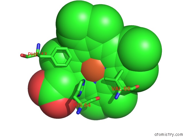
Mono view
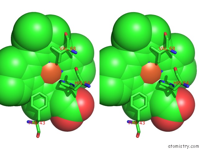
Stereo pair view
Mono view
Stereo pair view
A full contact list of Iron with other atoms in the Fe binding
site number 1 of Crystal Structure of Radiation-Induced Myoglobin Compound II - Intermediate H at pH 5.2 within 5.0Å range:
|
Reference:
H.-P.Hersleth,
T.Uchida,
A.K.Rohr,
T.Teschner,
V.Schunemann,
T.Kitagawa,
A.X.Trautwein,
C.H.Gorbitz,
K.K.Andersson.
Crystallographic and Spectroscopic Studies of Peroxide-Derived Myoglobin Compound II and Occurrence of Protonated Fe(IV)-O J.Biol.Chem. V. 282 23372 2007.
ISSN: ISSN 0021-9258
PubMed: 17565988
DOI: 10.1074/JBC.M701948200
Page generated: Thu Jul 17 04:06:56 2025
ISSN: ISSN 0021-9258
PubMed: 17565988
DOI: 10.1074/JBC.M701948200
Last articles
Fe in 2YXOFe in 2YRS
Fe in 2YXC
Fe in 2YNM
Fe in 2YVJ
Fe in 2YP1
Fe in 2YU2
Fe in 2YU1
Fe in 2YQB
Fe in 2YOO