Iron »
PDB 2v1j-2vlz »
2v1k »
Iron in PDB 2v1k: Crystal Structure of Ferrous Deoxymyoglobin at pH 6.8
Protein crystallography data
The structure of Crystal Structure of Ferrous Deoxymyoglobin at pH 6.8, PDB code: 2v1k
was solved by
H.-P.Hersleth,
C.H.Gorbitz,
K.K.Andersson,
with X-Ray Crystallography technique. A brief refinement statistics is given in the table below:
| Resolution Low / High (Å) | 26.78 / 1.25 |
| Space group | P 1 21 1 |
| Cell size a, b, c (Å), α, β, γ (°) | 62.900, 28.750, 35.440, 90.00, 105.72, 90.00 |
| R / Rfree (%) | 13.4 / 16.4 |
Iron Binding Sites:
The binding sites of Iron atom in the Crystal Structure of Ferrous Deoxymyoglobin at pH 6.8
(pdb code 2v1k). This binding sites where shown within
5.0 Angstroms radius around Iron atom.
In total only one binding site of Iron was determined in the Crystal Structure of Ferrous Deoxymyoglobin at pH 6.8, PDB code: 2v1k:
In total only one binding site of Iron was determined in the Crystal Structure of Ferrous Deoxymyoglobin at pH 6.8, PDB code: 2v1k:
Iron binding site 1 out of 1 in 2v1k
Go back to
Iron binding site 1 out
of 1 in the Crystal Structure of Ferrous Deoxymyoglobin at pH 6.8
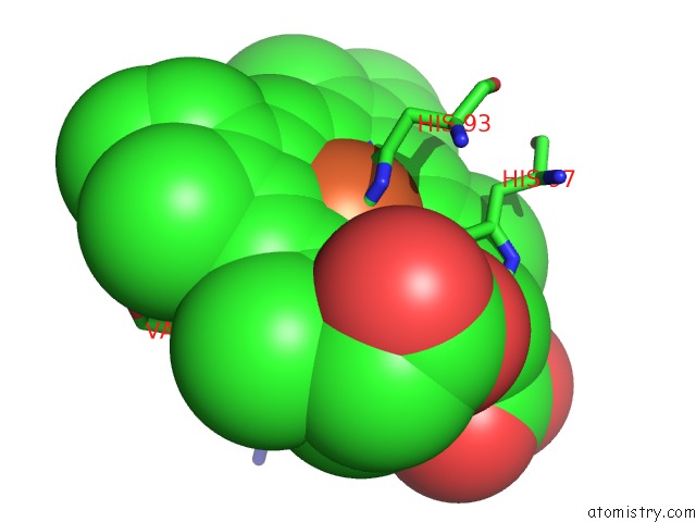
Mono view
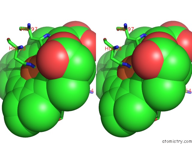
Stereo pair view
Mono view
Stereo pair view
A full contact list of Iron with other atoms in the Fe binding
site number 1 of Crystal Structure of Ferrous Deoxymyoglobin at pH 6.8 within 5.0Å range:
|
Reference:
H.-P.Hersleth,
T.Uchida,
A.K.Rohr,
T.Teschner,
V.Schunemann,
T.Kitagawa,
A.X.Trautwein,
C.H.Gorbitz,
K.K.Andersson.
Crystallographic and Spectroscopic Studies of Peroxide-Derived Myoglobin Compound II and Occurrence of Protonated Fe(IV)-O J.Biol.Chem. V. 282 23372 2007.
ISSN: ISSN 0021-9258
PubMed: 17565988
DOI: 10.1074/JBC.M701948200
Page generated: Sun Aug 4 02:33:07 2024
ISSN: ISSN 0021-9258
PubMed: 17565988
DOI: 10.1074/JBC.M701948200
Last articles
Zn in 9MJ5Zn in 9HNW
Zn in 9G0L
Zn in 9FNE
Zn in 9DZN
Zn in 9E0I
Zn in 9D32
Zn in 9DAK
Zn in 8ZXC
Zn in 8ZUF