Iron »
PDB 2v1j-2vlz »
2v45 »
Iron in PDB 2v45: A New Catalytic Mechanism of Periplasmic Nitrate Reductase From Desulfovibrio Desulfuricans Atcc 27774 From Crystallographic and Epr Data and Based on Detailed Analysis of the Sixth Ligand
Enzymatic activity of A New Catalytic Mechanism of Periplasmic Nitrate Reductase From Desulfovibrio Desulfuricans Atcc 27774 From Crystallographic and Epr Data and Based on Detailed Analysis of the Sixth Ligand
All present enzymatic activity of A New Catalytic Mechanism of Periplasmic Nitrate Reductase From Desulfovibrio Desulfuricans Atcc 27774 From Crystallographic and Epr Data and Based on Detailed Analysis of the Sixth Ligand:
1.7.99.4;
1.7.99.4;
Protein crystallography data
The structure of A New Catalytic Mechanism of Periplasmic Nitrate Reductase From Desulfovibrio Desulfuricans Atcc 27774 From Crystallographic and Epr Data and Based on Detailed Analysis of the Sixth Ligand, PDB code: 2v45
was solved by
S.Najmudin,
P.J.Gonzalez,
J.Trincao,
C.Coelho,
A.Mukhopadhyay,
C.C.Romao,
I.Moura,
J.J.Moura,
C.D.Brondino,
M.J.Romao,
with X-Ray Crystallography technique. A brief refinement statistics is given in the table below:
| Resolution Low / High (Å) | 91.67 / 2.40 |
| Space group | P 31 2 1 |
| Cell size a, b, c (Å), α, β, γ (°) | 105.902, 105.902, 130.241, 90.00, 90.00, 120.00 |
| R / Rfree (%) | 16.5 / 24 |
Other elements in 2v45:
The structure of A New Catalytic Mechanism of Periplasmic Nitrate Reductase From Desulfovibrio Desulfuricans Atcc 27774 From Crystallographic and Epr Data and Based on Detailed Analysis of the Sixth Ligand also contains other interesting chemical elements:
| Molybdenum | (Mo) | 1 atom |
| Chlorine | (Cl) | 1 atom |
Iron Binding Sites:
The binding sites of Iron atom in the A New Catalytic Mechanism of Periplasmic Nitrate Reductase From Desulfovibrio Desulfuricans Atcc 27774 From Crystallographic and Epr Data and Based on Detailed Analysis of the Sixth Ligand
(pdb code 2v45). This binding sites where shown within
5.0 Angstroms radius around Iron atom.
In total 4 binding sites of Iron where determined in the A New Catalytic Mechanism of Periplasmic Nitrate Reductase From Desulfovibrio Desulfuricans Atcc 27774 From Crystallographic and Epr Data and Based on Detailed Analysis of the Sixth Ligand, PDB code: 2v45:
Jump to Iron binding site number: 1; 2; 3; 4;
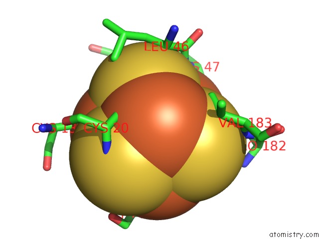
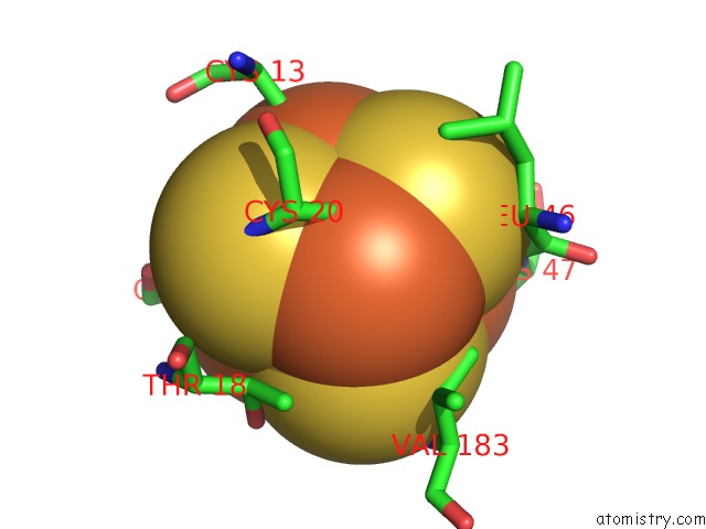
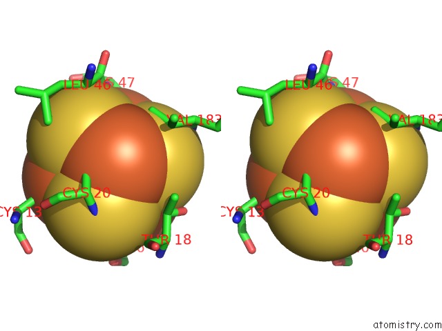
Iron binding site 1 out of 4 in 2v45
Go back to
Iron binding site 1 out
of 4 in the A New Catalytic Mechanism of Periplasmic Nitrate Reductase From Desulfovibrio Desulfuricans Atcc 27774 From Crystallographic and Epr Data and Based on Detailed Analysis of the Sixth Ligand
Mono view
Stereo pair view
Mono view
Stereo pair view
A full contact list of Iron with other atoms in the Fe binding
site number 1 of A New Catalytic Mechanism of Periplasmic Nitrate Reductase From Desulfovibrio Desulfuricans Atcc 27774 From Crystallographic and Epr Data and Based on Detailed Analysis of the Sixth Ligand within 5.0Å range:
|
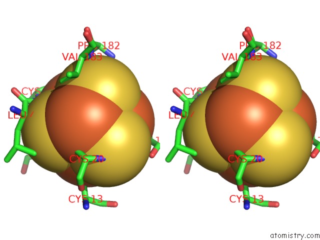
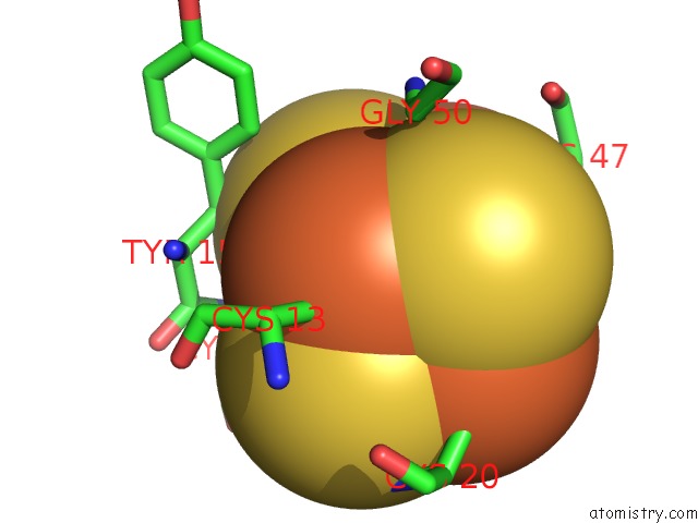
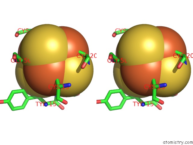
Iron binding site 2 out of 4 in 2v45
Go back to
Iron binding site 2 out
of 4 in the A New Catalytic Mechanism of Periplasmic Nitrate Reductase From Desulfovibrio Desulfuricans Atcc 27774 From Crystallographic and Epr Data and Based on Detailed Analysis of the Sixth Ligand
Mono view
Stereo pair view
Mono view
Stereo pair view
A full contact list of Iron with other atoms in the Fe binding
site number 2 of A New Catalytic Mechanism of Periplasmic Nitrate Reductase From Desulfovibrio Desulfuricans Atcc 27774 From Crystallographic and Epr Data and Based on Detailed Analysis of the Sixth Ligand within 5.0Å range:
|
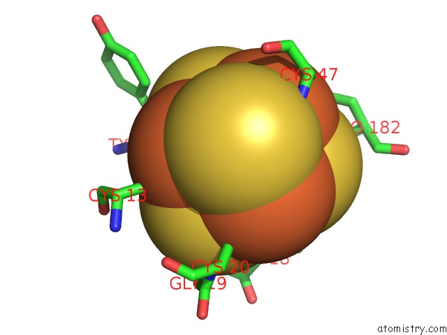
Iron binding site 3 out of 4 in 2v45
Go back to
Iron binding site 3 out
of 4 in the A New Catalytic Mechanism of Periplasmic Nitrate Reductase From Desulfovibrio Desulfuricans Atcc 27774 From Crystallographic and Epr Data and Based on Detailed Analysis of the Sixth Ligand
Mono view
Stereo pair view
Mono view
Stereo pair view
A full contact list of Iron with other atoms in the Fe binding
site number 3 of A New Catalytic Mechanism of Periplasmic Nitrate Reductase From Desulfovibrio Desulfuricans Atcc 27774 From Crystallographic and Epr Data and Based on Detailed Analysis of the Sixth Ligand within 5.0Å range:
|
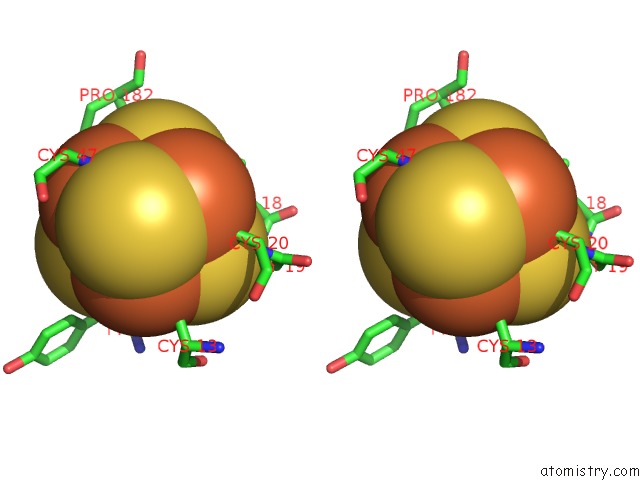
Iron binding site 4 out of 4 in 2v45
Go back to
Iron binding site 4 out
of 4 in the A New Catalytic Mechanism of Periplasmic Nitrate Reductase From Desulfovibrio Desulfuricans Atcc 27774 From Crystallographic and Epr Data and Based on Detailed Analysis of the Sixth Ligand
Mono view
Stereo pair view
Mono view
Stereo pair view
A full contact list of Iron with other atoms in the Fe binding
site number 4 of A New Catalytic Mechanism of Periplasmic Nitrate Reductase From Desulfovibrio Desulfuricans Atcc 27774 From Crystallographic and Epr Data and Based on Detailed Analysis of the Sixth Ligand within 5.0Å range:
|
Reference:
S.Najmudin,
P.J.Gonzalez,
J.Trincao,
C.Coelho,
A.Mukhopadhyay,
N.M.F.S.A.Cerqueira,
C.C.Romao,
I.Moura,
J.J.G.Moura,
C.D.Brondino,
M.J.Romao.
Periplasmic Nitrate Reductase Revisited: A Sulfur Atom Completes the Sixth Coordination of the Catalytic Molybdenum. J.Biol.Inorg.Chem. V. 13 737 2008.
ISSN: ISSN 0949-8257
PubMed: 18327621
DOI: 10.1007/S00775-008-0359-6
Page generated: Sun Aug 4 02:33:07 2024
ISSN: ISSN 0949-8257
PubMed: 18327621
DOI: 10.1007/S00775-008-0359-6
Last articles
Cl in 5VUSCl in 5VTK
Cl in 5VSV
Cl in 5VR0
Cl in 5VTI
Cl in 5VTD
Cl in 5VT7
Cl in 5VSK
Cl in 5VSB
Cl in 5VS7