Iron »
PDB 2v1j-2vlz »
2vcs »
Iron in PDB 2vcs: Structure of Isoniazid (Inh) Bound to Cytosolic Soybean Ascorbate Peroxidase Mutant H42A
Enzymatic activity of Structure of Isoniazid (Inh) Bound to Cytosolic Soybean Ascorbate Peroxidase Mutant H42A
All present enzymatic activity of Structure of Isoniazid (Inh) Bound to Cytosolic Soybean Ascorbate Peroxidase Mutant H42A:
1.11.1.11;
1.11.1.11;
Protein crystallography data
The structure of Structure of Isoniazid (Inh) Bound to Cytosolic Soybean Ascorbate Peroxidase Mutant H42A, PDB code: 2vcs
was solved by
C.L.Metcalfe,
I.K.Macdonald,
K.A.Brown,
E.L.Raven,
P.C.E.Moody,
with X-Ray Crystallography technique. A brief refinement statistics is given in the table below:
| Resolution Low / High (Å) | 27.62 / 1.68 |
| Space group | P 42 21 2 |
| Cell size a, b, c (Å), α, β, γ (°) | 81.735, 81.735, 74.937, 90.00, 90.00, 90.00 |
| R / Rfree (%) | 16.6 / 21 |
Iron Binding Sites:
The binding sites of Iron atom in the Structure of Isoniazid (Inh) Bound to Cytosolic Soybean Ascorbate Peroxidase Mutant H42A
(pdb code 2vcs). This binding sites where shown within
5.0 Angstroms radius around Iron atom.
In total only one binding site of Iron was determined in the Structure of Isoniazid (Inh) Bound to Cytosolic Soybean Ascorbate Peroxidase Mutant H42A, PDB code: 2vcs:
In total only one binding site of Iron was determined in the Structure of Isoniazid (Inh) Bound to Cytosolic Soybean Ascorbate Peroxidase Mutant H42A, PDB code: 2vcs:
Iron binding site 1 out of 1 in 2vcs
Go back to
Iron binding site 1 out
of 1 in the Structure of Isoniazid (Inh) Bound to Cytosolic Soybean Ascorbate Peroxidase Mutant H42A
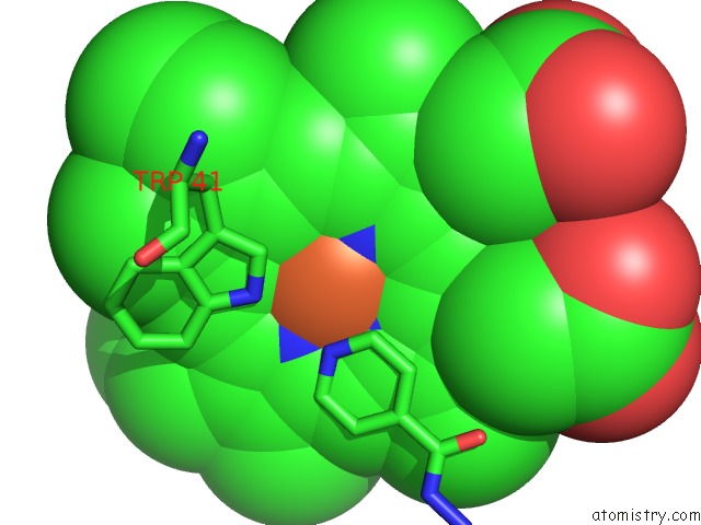
Mono view
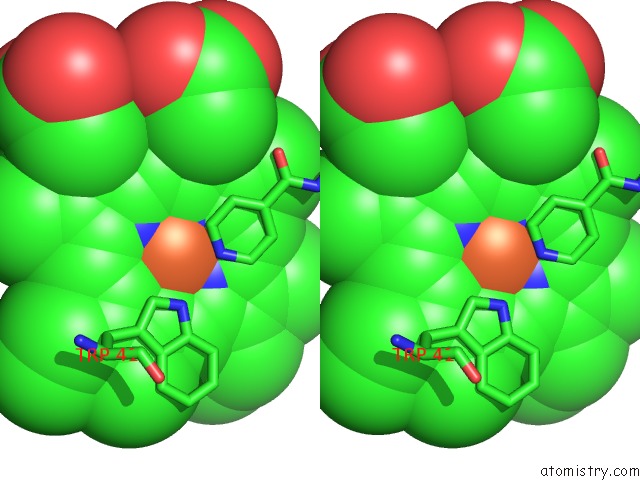
Stereo pair view
Mono view
Stereo pair view
A full contact list of Iron with other atoms in the Fe binding
site number 1 of Structure of Isoniazid (Inh) Bound to Cytosolic Soybean Ascorbate Peroxidase Mutant H42A within 5.0Å range:
|
Reference:
C.L.Metcalfe,
I.K.Macdonald,
E.J.Murphy,
K.A.Brown,
E.L.Raven,
P.C.E.Moody.
The Tuberculosis Prodrug Isoniazid Bound to Activating Peroxidases. J.Biol.Chem. V. 283 6193 2008.
ISSN: ISSN 0021-9258
PubMed: 18056997
DOI: 10.1074/JBC.M707412200
Page generated: Sun Aug 4 02:36:00 2024
ISSN: ISSN 0021-9258
PubMed: 18056997
DOI: 10.1074/JBC.M707412200
Last articles
Zn in 9MJ5Zn in 9HNW
Zn in 9G0L
Zn in 9FNE
Zn in 9DZN
Zn in 9E0I
Zn in 9D32
Zn in 9DAK
Zn in 8ZXC
Zn in 8ZUF