Iron »
PDB 2v1j-2vlz »
2vdc »
Iron in PDB 2vdc: The 9.5 A Resolution Structure of Glutamate Synthase From Cryo- Electron Microscopy and Its Oligomerization Behavior in Solution: Functional Implications.
Enzymatic activity of The 9.5 A Resolution Structure of Glutamate Synthase From Cryo- Electron Microscopy and Its Oligomerization Behavior in Solution: Functional Implications.
All present enzymatic activity of The 9.5 A Resolution Structure of Glutamate Synthase From Cryo- Electron Microscopy and Its Oligomerization Behavior in Solution: Functional Implications.:
1.4.1.13;
1.4.1.13;
Iron Binding Sites:
Pages:
>>> Page 1 <<< Page 2, Binding sites: 11 - 20; Page 3, Binding sites: 21 - 30; Page 4, Binding sites: 31 - 40; Page 5, Binding sites: 41 - 50; Page 6, Binding sites: 51 - 60; Page 7, Binding sites: 61 - 66;Binding sites:
The binding sites of Iron atom in the The 9.5 A Resolution Structure of Glutamate Synthase From Cryo- Electron Microscopy and Its Oligomerization Behavior in Solution: Functional Implications. (pdb code 2vdc). This binding sites where shown within 5.0 Angstroms radius around Iron atom.In total 66 binding sites of Iron where determined in the The 9.5 A Resolution Structure of Glutamate Synthase From Cryo- Electron Microscopy and Its Oligomerization Behavior in Solution: Functional Implications., PDB code: 2vdc:
Jump to Iron binding site number: 1; 2; 3; 4; 5; 6; 7; 8; 9; 10;
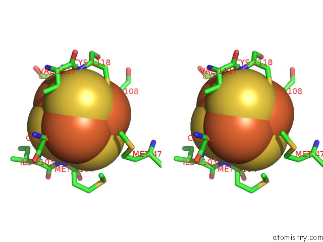
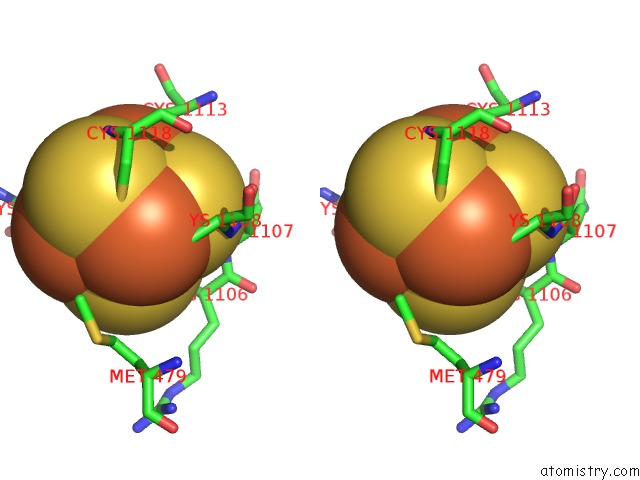
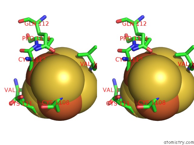
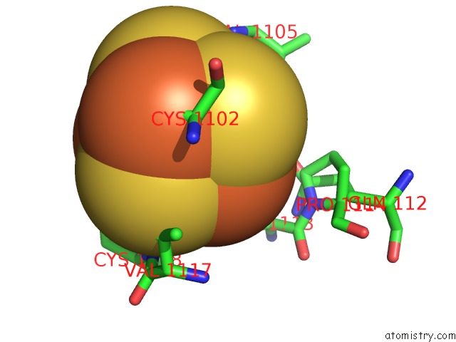
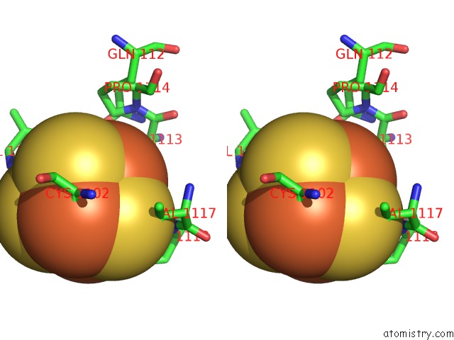
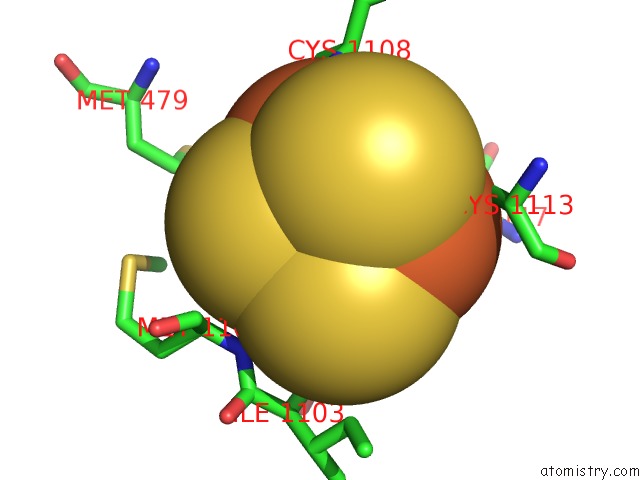
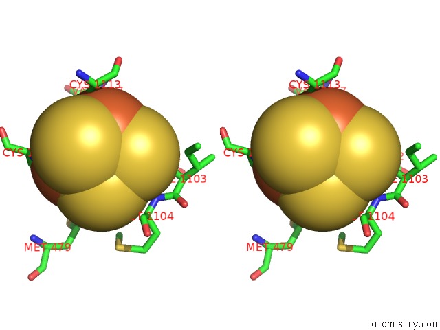
Iron binding site 1 out of 66 in 2vdc
Go back to
Iron binding site 1 out
of 66 in the The 9.5 A Resolution Structure of Glutamate Synthase From Cryo- Electron Microscopy and Its Oligomerization Behavior in Solution: Functional Implications.

Mono view
Stereo pair view
Mono view
Stereo pair view
A full contact list of Iron with other atoms in the Fe binding
site number 1 of The 9.5 A Resolution Structure of Glutamate Synthase From Cryo- Electron Microscopy and Its Oligomerization Behavior in Solution: Functional Implications. within 5.0Å range:
|
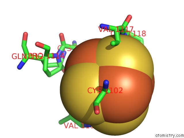
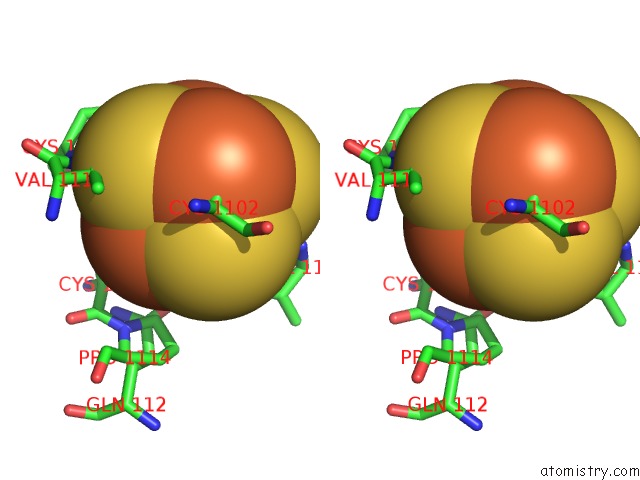
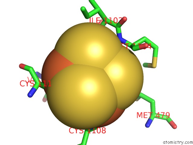
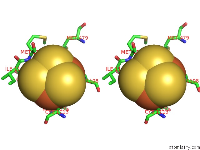
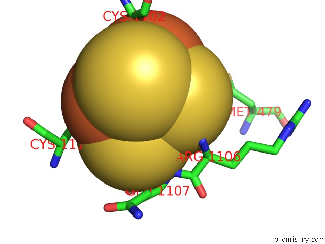
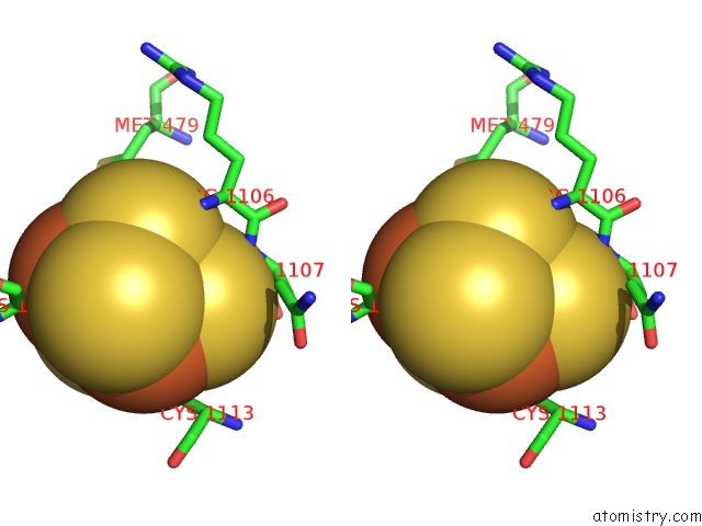
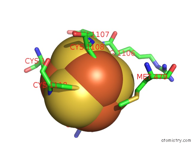
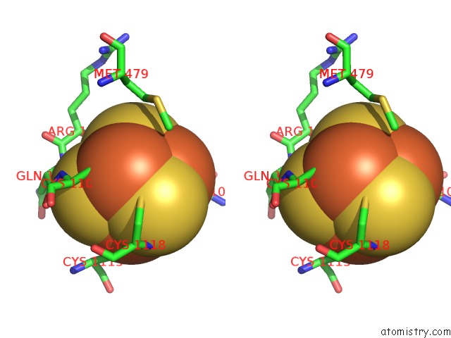
Iron binding site 2 out of 66 in 2vdc
Go back to
Iron binding site 2 out
of 66 in the The 9.5 A Resolution Structure of Glutamate Synthase From Cryo- Electron Microscopy and Its Oligomerization Behavior in Solution: Functional Implications.
Mono view
Stereo pair view
Mono view
Stereo pair view
A full contact list of Iron with other atoms in the Fe binding
site number 2 of The 9.5 A Resolution Structure of Glutamate Synthase From Cryo- Electron Microscopy and Its Oligomerization Behavior in Solution: Functional Implications. within 5.0Å range:
|
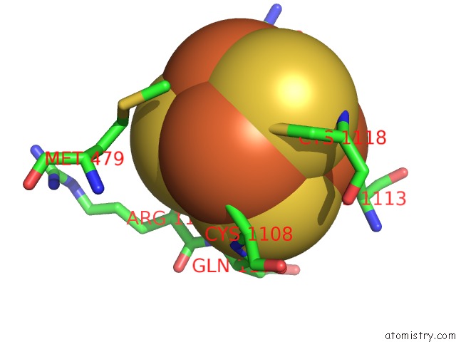
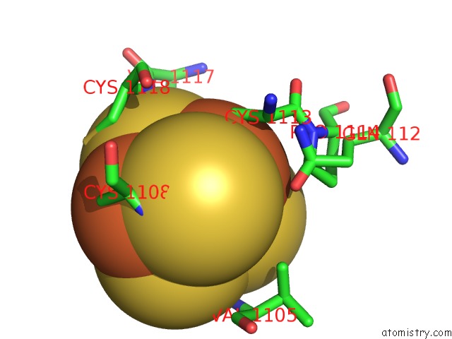
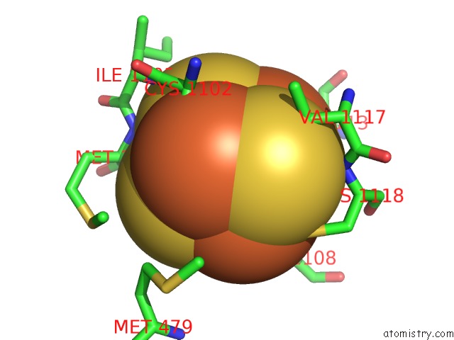
Iron binding site 3 out of 66 in 2vdc
Go back to
Iron binding site 3 out
of 66 in the The 9.5 A Resolution Structure of Glutamate Synthase From Cryo- Electron Microscopy and Its Oligomerization Behavior in Solution: Functional Implications.
Mono view
Stereo pair view
Mono view
Stereo pair view
A full contact list of Iron with other atoms in the Fe binding
site number 3 of The 9.5 A Resolution Structure of Glutamate Synthase From Cryo- Electron Microscopy and Its Oligomerization Behavior in Solution: Functional Implications. within 5.0Å range:
|
Iron binding site 4 out of 66 in 2vdc
Go back to
Iron binding site 4 out
of 66 in the The 9.5 A Resolution Structure of Glutamate Synthase From Cryo- Electron Microscopy and Its Oligomerization Behavior in Solution: Functional Implications.
Mono view
Stereo pair view
Mono view
Stereo pair view
A full contact list of Iron with other atoms in the Fe binding
site number 4 of The 9.5 A Resolution Structure of Glutamate Synthase From Cryo- Electron Microscopy and Its Oligomerization Behavior in Solution: Functional Implications. within 5.0Å range:
|
Iron binding site 5 out of 66 in 2vdc
Go back to
Iron binding site 5 out
of 66 in the The 9.5 A Resolution Structure of Glutamate Synthase From Cryo- Electron Microscopy and Its Oligomerization Behavior in Solution: Functional Implications.
Mono view
Stereo pair view
Mono view
Stereo pair view
A full contact list of Iron with other atoms in the Fe binding
site number 5 of The 9.5 A Resolution Structure of Glutamate Synthase From Cryo- Electron Microscopy and Its Oligomerization Behavior in Solution: Functional Implications. within 5.0Å range:
|
Iron binding site 6 out of 66 in 2vdc
Go back to
Iron binding site 6 out
of 66 in the The 9.5 A Resolution Structure of Glutamate Synthase From Cryo- Electron Microscopy and Its Oligomerization Behavior in Solution: Functional Implications.
Mono view
Stereo pair view
Mono view
Stereo pair view
A full contact list of Iron with other atoms in the Fe binding
site number 6 of The 9.5 A Resolution Structure of Glutamate Synthase From Cryo- Electron Microscopy and Its Oligomerization Behavior in Solution: Functional Implications. within 5.0Å range:
|
Iron binding site 7 out of 66 in 2vdc
Go back to
Iron binding site 7 out
of 66 in the The 9.5 A Resolution Structure of Glutamate Synthase From Cryo- Electron Microscopy and Its Oligomerization Behavior in Solution: Functional Implications.
Mono view

Stereo pair view
Mono view
Stereo pair view
A full contact list of Iron with other atoms in the Fe binding
site number 7 of The 9.5 A Resolution Structure of Glutamate Synthase From Cryo- Electron Microscopy and Its Oligomerization Behavior in Solution: Functional Implications. within 5.0Å range:
|
Iron binding site 8 out of 66 in 2vdc
Go back to
Iron binding site 8 out
of 66 in the The 9.5 A Resolution Structure of Glutamate Synthase From Cryo- Electron Microscopy and Its Oligomerization Behavior in Solution: Functional Implications.
Mono view
Stereo pair view
Mono view
Stereo pair view
A full contact list of Iron with other atoms in the Fe binding
site number 8 of The 9.5 A Resolution Structure of Glutamate Synthase From Cryo- Electron Microscopy and Its Oligomerization Behavior in Solution: Functional Implications. within 5.0Å range:
|
Iron binding site 9 out of 66 in 2vdc
Go back to
Iron binding site 9 out
of 66 in the The 9.5 A Resolution Structure of Glutamate Synthase From Cryo- Electron Microscopy and Its Oligomerization Behavior in Solution: Functional Implications.
Mono view
Stereo pair view
Mono view
Stereo pair view
A full contact list of Iron with other atoms in the Fe binding
site number 9 of The 9.5 A Resolution Structure of Glutamate Synthase From Cryo- Electron Microscopy and Its Oligomerization Behavior in Solution: Functional Implications. within 5.0Å range:
|
Iron binding site 10 out of 66 in 2vdc
Go back to
Iron binding site 10 out
of 66 in the The 9.5 A Resolution Structure of Glutamate Synthase From Cryo- Electron Microscopy and Its Oligomerization Behavior in Solution: Functional Implications.
Mono view
Stereo pair view
Mono view
Stereo pair view
A full contact list of Iron with other atoms in the Fe binding
site number 10 of The 9.5 A Resolution Structure of Glutamate Synthase From Cryo- Electron Microscopy and Its Oligomerization Behavior in Solution: Functional Implications. within 5.0Å range:
|
Reference:
M.Cottevieille,
E.Larquet,
S.Jonic,
M.V.Petoukhov,
G.Caprini,
S.Paravisi,
D.I.Svergun,
M.A.Vanoni,
N.Boisset.
The Subnanometer Resolution Structure of the Glutamate Synthase 1.2-Mda Hexamer By Cryoelectron Microscopy and Its Oligomerization Behavior in Solution: Functional Implications. J.Biol.Chem. V. 283 8237 2008.
ISSN: ISSN 0021-9258
PubMed: 18199747
DOI: 10.1074/JBC.M708529200
Page generated: Sun Aug 4 02:37:13 2024
ISSN: ISSN 0021-9258
PubMed: 18199747
DOI: 10.1074/JBC.M708529200
Last articles
Zn in 9MJ5Zn in 9HNW
Zn in 9G0L
Zn in 9FNE
Zn in 9DZN
Zn in 9E0I
Zn in 9D32
Zn in 9DAK
Zn in 8ZXC
Zn in 8ZUF