Iron »
PDB 2v1j-2vlz »
2veb »
Iron in PDB 2veb: High Resolution Structure of Protoglobin From Methanosarcina Acetivorans C2A
Protein crystallography data
The structure of High Resolution Structure of Protoglobin From Methanosarcina Acetivorans C2A, PDB code: 2veb
was solved by
M.Nardini,
A.Pesce,
L.Thijs,
J.A.Saito,
S.Dewilde,
M.Alam,
P.Ascenzi,
M.Coletta,
C.Ciaccio,
L.Moens,
M.Bolognesi,
with X-Ray Crystallography technique. A brief refinement statistics is given in the table below:
| Resolution Low / High (Å) | 51.43 / 1.30 |
| Space group | C 1 2 1 |
| Cell size a, b, c (Å), α, β, γ (°) | 80.059, 49.271, 51.513, 90.00, 92.88, 90.00 |
| R / Rfree (%) | 16 / 19 |
Iron Binding Sites:
The binding sites of Iron atom in the High Resolution Structure of Protoglobin From Methanosarcina Acetivorans C2A
(pdb code 2veb). This binding sites where shown within
5.0 Angstroms radius around Iron atom.
In total only one binding site of Iron was determined in the High Resolution Structure of Protoglobin From Methanosarcina Acetivorans C2A, PDB code: 2veb:
In total only one binding site of Iron was determined in the High Resolution Structure of Protoglobin From Methanosarcina Acetivorans C2A, PDB code: 2veb:
Iron binding site 1 out of 1 in 2veb
Go back to
Iron binding site 1 out
of 1 in the High Resolution Structure of Protoglobin From Methanosarcina Acetivorans C2A
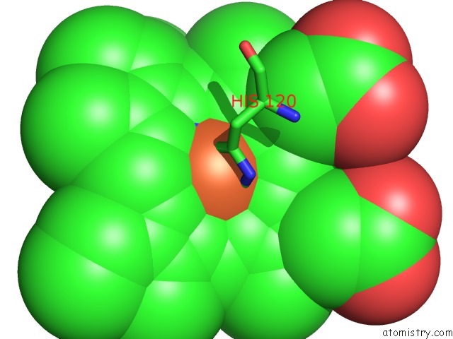
Mono view
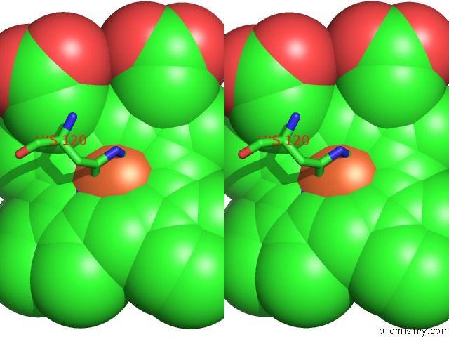
Stereo pair view
Mono view
Stereo pair view
A full contact list of Iron with other atoms in the Fe binding
site number 1 of High Resolution Structure of Protoglobin From Methanosarcina Acetivorans C2A within 5.0Å range:
|
Reference:
M.Nardini,
A.Pesce,
L.Thijs,
J.A.Saito,
S.Dewilde,
M.Alam,
P.Ascenzi,
M.Coletta,
C.Ciaccio,
L.Moens,
M.Bolognesi.
Archaeal Protoglobin Structure Indicates New Ligand Diffusion Paths and Modulation of Haem- Reactivity. Embo Rep. V. 9 157 2008.
ISSN: ISSN 1469-221X
PubMed: 18188182
DOI: 10.1038/SJ.EMBOR.7401153
Page generated: Sun Aug 4 02:37:27 2024
ISSN: ISSN 1469-221X
PubMed: 18188182
DOI: 10.1038/SJ.EMBOR.7401153
Last articles
Zn in 9MJ5Zn in 9HNW
Zn in 9G0L
Zn in 9FNE
Zn in 9DZN
Zn in 9E0I
Zn in 9D32
Zn in 9DAK
Zn in 8ZXC
Zn in 8ZUF