Iron »
PDB 2v1j-2vlz »
2vhd »
Iron in PDB 2vhd: Crystal Structure of the Di-Haem Cytochrome C Peroxidase From Pseudomonas Aeruginosa - Mixed Valence Form
Enzymatic activity of Crystal Structure of the Di-Haem Cytochrome C Peroxidase From Pseudomonas Aeruginosa - Mixed Valence Form
All present enzymatic activity of Crystal Structure of the Di-Haem Cytochrome C Peroxidase From Pseudomonas Aeruginosa - Mixed Valence Form:
1.11.1.5;
1.11.1.5;
Protein crystallography data
The structure of Crystal Structure of the Di-Haem Cytochrome C Peroxidase From Pseudomonas Aeruginosa - Mixed Valence Form, PDB code: 2vhd
was solved by
A.Echalier,
T.Brittain,
J.Wright,
S.Boycheva,
G.B.Mortuza,
V.Fulop,
N.J.Watmough,
with X-Ray Crystallography technique. A brief refinement statistics is given in the table below:
| Resolution Low / High (Å) | 76.70 / 2.30 |
| Space group | C 1 2 1 |
| Cell size a, b, c (Å), α, β, γ (°) | 173.200, 44.800, 106.200, 90.00, 106.70, 90.00 |
| R / Rfree (%) | 18.4 / 24.6 |
Other elements in 2vhd:
The structure of Crystal Structure of the Di-Haem Cytochrome C Peroxidase From Pseudomonas Aeruginosa - Mixed Valence Form also contains other interesting chemical elements:
| Calcium | (Ca) | 2 atoms |
Iron Binding Sites:
The binding sites of Iron atom in the Crystal Structure of the Di-Haem Cytochrome C Peroxidase From Pseudomonas Aeruginosa - Mixed Valence Form
(pdb code 2vhd). This binding sites where shown within
5.0 Angstroms radius around Iron atom.
In total 4 binding sites of Iron where determined in the Crystal Structure of the Di-Haem Cytochrome C Peroxidase From Pseudomonas Aeruginosa - Mixed Valence Form, PDB code: 2vhd:
Jump to Iron binding site number: 1; 2; 3; 4;
In total 4 binding sites of Iron where determined in the Crystal Structure of the Di-Haem Cytochrome C Peroxidase From Pseudomonas Aeruginosa - Mixed Valence Form, PDB code: 2vhd:
Jump to Iron binding site number: 1; 2; 3; 4;
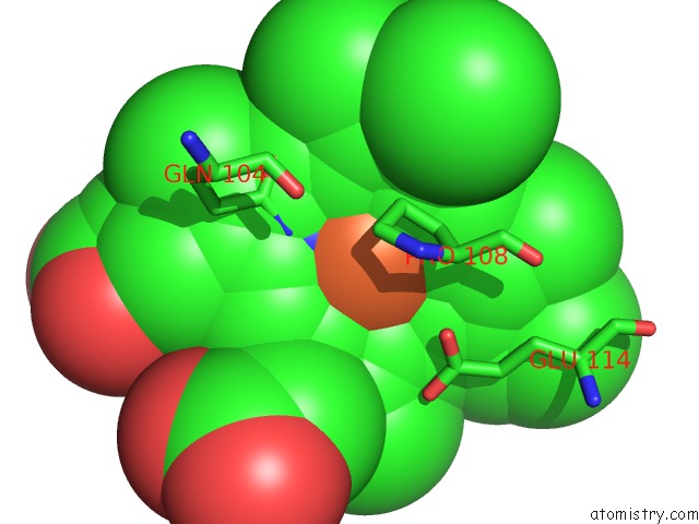
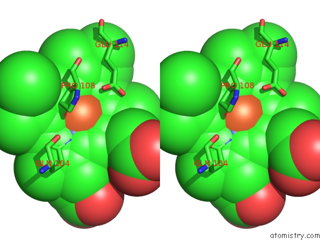
Iron binding site 1 out of 4 in 2vhd
Go back to
Iron binding site 1 out
of 4 in the Crystal Structure of the Di-Haem Cytochrome C Peroxidase From Pseudomonas Aeruginosa - Mixed Valence Form
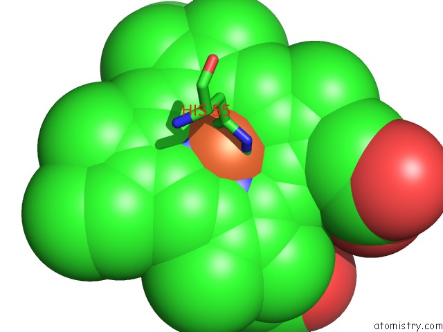
Mono view
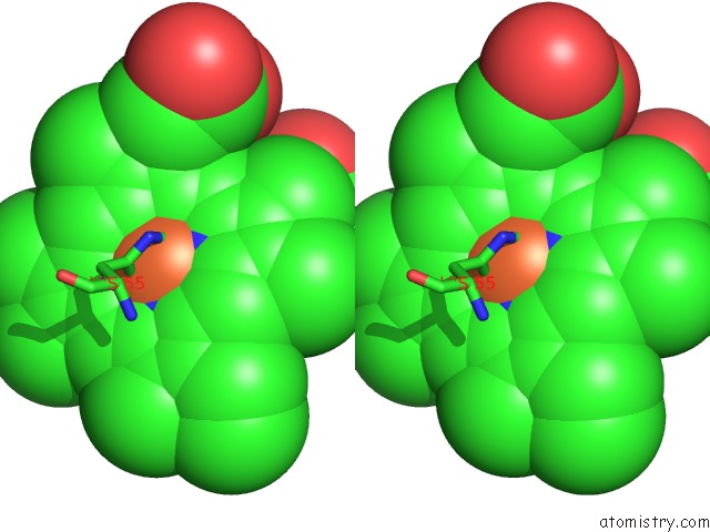
Stereo pair view
Mono view
Stereo pair view
A full contact list of Iron with other atoms in the Fe binding
site number 1 of Crystal Structure of the Di-Haem Cytochrome C Peroxidase From Pseudomonas Aeruginosa - Mixed Valence Form within 5.0Å range:
|
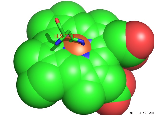
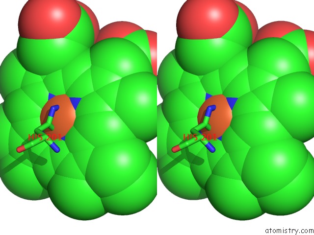
Iron binding site 2 out of 4 in 2vhd
Go back to
Iron binding site 2 out
of 4 in the Crystal Structure of the Di-Haem Cytochrome C Peroxidase From Pseudomonas Aeruginosa - Mixed Valence Form
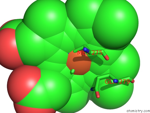
Mono view
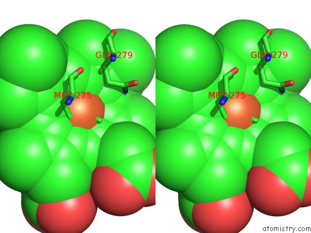
Stereo pair view
Mono view
Stereo pair view
A full contact list of Iron with other atoms in the Fe binding
site number 2 of Crystal Structure of the Di-Haem Cytochrome C Peroxidase From Pseudomonas Aeruginosa - Mixed Valence Form within 5.0Å range:
|
Iron binding site 3 out of 4 in 2vhd
Go back to
Iron binding site 3 out
of 4 in the Crystal Structure of the Di-Haem Cytochrome C Peroxidase From Pseudomonas Aeruginosa - Mixed Valence Form
Mono view
Stereo pair view
Mono view
Stereo pair view
A full contact list of Iron with other atoms in the Fe binding
site number 3 of Crystal Structure of the Di-Haem Cytochrome C Peroxidase From Pseudomonas Aeruginosa - Mixed Valence Form within 5.0Å range:
|
Iron binding site 4 out of 4 in 2vhd
Go back to
Iron binding site 4 out
of 4 in the Crystal Structure of the Di-Haem Cytochrome C Peroxidase From Pseudomonas Aeruginosa - Mixed Valence Form
Mono view
Stereo pair view
Mono view
Stereo pair view
A full contact list of Iron with other atoms in the Fe binding
site number 4 of Crystal Structure of the Di-Haem Cytochrome C Peroxidase From Pseudomonas Aeruginosa - Mixed Valence Form within 5.0Å range:
|
Reference:
A.Echalier,
T.Brittain,
J.Wright,
S.Boycheva,
G.B.Mortuza,
V.Fulop,
N.J.Watmough.
Redox-Linked Structural Changes Associated with the Formation of A Catalytically Competent Form of the Diheme Cytochrome C Peroxidase From Pseudomonas Aeruginosa Biochemistry V. 47 1947 2008.
ISSN: ISSN 0006-2960
PubMed: 18217775
DOI: 10.1021/BI702064F
Page generated: Sun Aug 4 02:38:40 2024
ISSN: ISSN 0006-2960
PubMed: 18217775
DOI: 10.1021/BI702064F
Last articles
Zn in 9J0NZn in 9J0O
Zn in 9J0P
Zn in 9FJX
Zn in 9EKB
Zn in 9C0F
Zn in 9CAH
Zn in 9CH0
Zn in 9CH3
Zn in 9CH1