Iron »
PDB 2v1j-2vlz »
2vhl »
Iron in PDB 2vhl: The Three-Dimensional Structure of the N-Acetylglucosamine- 6-Phosphate Deacetylase From Bacillus Subtilis
Enzymatic activity of The Three-Dimensional Structure of the N-Acetylglucosamine- 6-Phosphate Deacetylase From Bacillus Subtilis
All present enzymatic activity of The Three-Dimensional Structure of the N-Acetylglucosamine- 6-Phosphate Deacetylase From Bacillus Subtilis:
3.5.1.25;
3.5.1.25;
Protein crystallography data
The structure of The Three-Dimensional Structure of the N-Acetylglucosamine- 6-Phosphate Deacetylase From Bacillus Subtilis, PDB code: 2vhl
was solved by
F.Vincent,
D.Yates,
E.Garman,
G.J.Davies,
with X-Ray Crystallography technique. A brief refinement statistics is given in the table below:
| Resolution Low / High (Å) | 21.84 / 2.05 |
| Space group | P 21 21 2 |
| Cell size a, b, c (Å), α, β, γ (°) | 51.680, 107.740, 188.250, 90.00, 90.00, 90.00 |
| R / Rfree (%) | 20.5 / 26 |
Iron Binding Sites:
The binding sites of Iron atom in the The Three-Dimensional Structure of the N-Acetylglucosamine- 6-Phosphate Deacetylase From Bacillus Subtilis
(pdb code 2vhl). This binding sites where shown within
5.0 Angstroms radius around Iron atom.
In total 4 binding sites of Iron where determined in the The Three-Dimensional Structure of the N-Acetylglucosamine- 6-Phosphate Deacetylase From Bacillus Subtilis, PDB code: 2vhl:
Jump to Iron binding site number: 1; 2; 3; 4;
In total 4 binding sites of Iron where determined in the The Three-Dimensional Structure of the N-Acetylglucosamine- 6-Phosphate Deacetylase From Bacillus Subtilis, PDB code: 2vhl:
Jump to Iron binding site number: 1; 2; 3; 4;
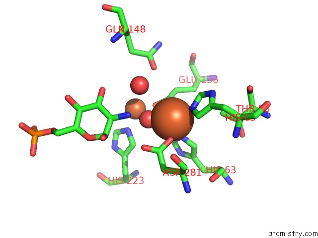
Iron binding site 1 out of 4 in 2vhl
Go back to
Iron binding site 1 out
of 4 in the The Three-Dimensional Structure of the N-Acetylglucosamine- 6-Phosphate Deacetylase From Bacillus Subtilis
Mono view
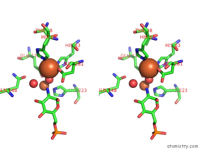
Stereo pair view
Mono view
Stereo pair view
A full contact list of Iron with other atoms in the Fe binding
site number 1 of The Three-Dimensional Structure of the N-Acetylglucosamine- 6-Phosphate Deacetylase From Bacillus Subtilis within 5.0Å range:
|
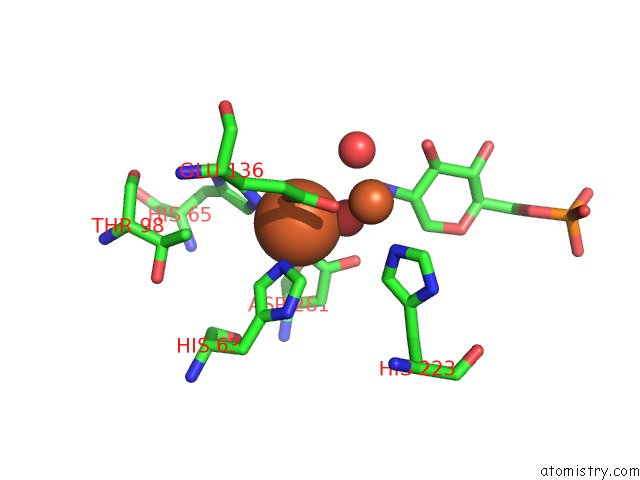
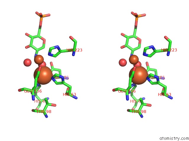
Iron binding site 2 out of 4 in 2vhl
Go back to
Iron binding site 2 out
of 4 in the The Three-Dimensional Structure of the N-Acetylglucosamine- 6-Phosphate Deacetylase From Bacillus Subtilis

Mono view

Stereo pair view
Mono view
Stereo pair view
A full contact list of Iron with other atoms in the Fe binding
site number 2 of The Three-Dimensional Structure of the N-Acetylglucosamine- 6-Phosphate Deacetylase From Bacillus Subtilis within 5.0Å range:
|
Iron binding site 3 out of 4 in 2vhl
Go back to
Iron binding site 3 out
of 4 in the The Three-Dimensional Structure of the N-Acetylglucosamine- 6-Phosphate Deacetylase From Bacillus Subtilis

Mono view

Stereo pair view
Mono view
Stereo pair view
A full contact list of Iron with other atoms in the Fe binding
site number 3 of The Three-Dimensional Structure of the N-Acetylglucosamine- 6-Phosphate Deacetylase From Bacillus Subtilis within 5.0Å range:
|
Iron binding site 4 out of 4 in 2vhl
Go back to
Iron binding site 4 out
of 4 in the The Three-Dimensional Structure of the N-Acetylglucosamine- 6-Phosphate Deacetylase From Bacillus Subtilis
Mono view
Stereo pair view
Mono view
Stereo pair view
A full contact list of Iron with other atoms in the Fe binding
site number 4 of The Three-Dimensional Structure of the N-Acetylglucosamine- 6-Phosphate Deacetylase From Bacillus Subtilis within 5.0Å range:
|
Reference:
F.Vincent,
D.Yates,
E.Garman,
G.J.Davies.
The Three-Dimensional Structure of the N- Acetylglucosamine-6-Phosphate Deacetylase From Bacillus Subtilis J.Biol.Chem. V. 279 2809 2004.
ISSN: ISSN 0021-9258
PubMed: 14557261
DOI: 10.1074/JBC.M310165200
Page generated: Sun Aug 4 02:38:47 2024
ISSN: ISSN 0021-9258
PubMed: 14557261
DOI: 10.1074/JBC.M310165200
Last articles
F in 7N33F in 7N4Q
F in 7N4N
F in 7N2A
F in 7MXY
F in 7MYY
F in 7N13
F in 7MYU
F in 7MYR
F in 7MYO