Iron »
PDB 2v1j-2vlz »
2vlx »
Iron in PDB 2vlx: Crystal Structure of Peroxymyoglobin Generated By Cryoradiolytic Reduction of Myoglobin Compound III
Protein crystallography data
The structure of Crystal Structure of Peroxymyoglobin Generated By Cryoradiolytic Reduction of Myoglobin Compound III, PDB code: 2vlx
was solved by
H.-P.Hersleth,
C.H.Gorbitz,
K.K.Andersson,
with X-Ray Crystallography technique. A brief refinement statistics is given in the table below:
| Resolution Low / High (Å) | 26.60 / 1.30 |
| Space group | P 1 21 1 |
| Cell size a, b, c (Å), α, β, γ (°) | 63.617, 28.663, 35.271, 90.00, 105.85, 90.00 |
| R / Rfree (%) | 14.7 / 17.5 |
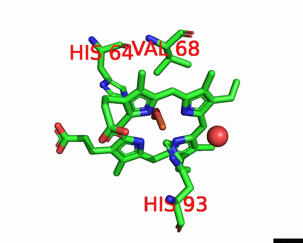
Iron Binding Sites:
The binding sites of Iron atom in the Crystal Structure of Peroxymyoglobin Generated By Cryoradiolytic Reduction of Myoglobin Compound III
(pdb code 2vlx). This binding sites where shown within
5.0 Angstroms radius around Iron atom.
In total only one binding site of Iron was determined in the Crystal Structure of Peroxymyoglobin Generated By Cryoradiolytic Reduction of Myoglobin Compound III, PDB code: 2vlx:
In total only one binding site of Iron was determined in the Crystal Structure of Peroxymyoglobin Generated By Cryoradiolytic Reduction of Myoglobin Compound III, PDB code: 2vlx:
Iron binding site 1 out of 1 in 2vlx
Go back to
Iron binding site 1 out
of 1 in the Crystal Structure of Peroxymyoglobin Generated By Cryoradiolytic Reduction of Myoglobin Compound III
Mono view

Stereo pair view
Mono view
Stereo pair view
A full contact list of Iron with other atoms in the Fe binding
site number 1 of Crystal Structure of Peroxymyoglobin Generated By Cryoradiolytic Reduction of Myoglobin Compound III within 5.0Å range:
|
Reference:
H.-P.Hersleth,
Y.-W.Hsiao,
U.Ryde,
C.H.Gorbitz,
K.K.Andersson.
The Crystal Structure of Peroxymyoglobin Generated Through Cryoradiolytic Reduction of Myoglobin Compound III During Data Collection. Biochem.J. V. 412 257 2008.
ISSN: ISSN 0264-6021
PubMed: 18215120
DOI: 10.1042/BJ20070921
Page generated: Thu Jul 17 04:18:10 2025
ISSN: ISSN 0264-6021
PubMed: 18215120
DOI: 10.1042/BJ20070921
Last articles
Fe in 2YXOFe in 2YRS
Fe in 2YXC
Fe in 2YNM
Fe in 2YVJ
Fe in 2YP1
Fe in 2YU2
Fe in 2YU1
Fe in 2YQB
Fe in 2YOO