Iron »
PDB 2w3h-2wl3 »
2w6v »
Iron in PDB 2w6v: Structure of Human Deoxy Hemoglobin A in Complex with Xenon
Protein crystallography data
The structure of Structure of Human Deoxy Hemoglobin A in Complex with Xenon, PDB code: 2w6v
was solved by
A.E.Miele,
F.Draghi,
G.Sciara,
K.A.Johnson,
F.Renzi,
B.Vallone,
M.Brunori,
C.Savino,
with X-Ray Crystallography technique. A brief refinement statistics is given in the table below:
| Resolution Low / High (Å) | 22.90 / 1.80 |
| Space group | P 1 21 1 |
| Cell size a, b, c (Å), α, β, γ (°) | 62.645, 82.328, 53.552, 90.00, 99.79, 90.00 |
| R / Rfree (%) | 16.2 / 21.6 |
Other elements in 2w6v:
The structure of Structure of Human Deoxy Hemoglobin A in Complex with Xenon also contains other interesting chemical elements:
| Xenon | (Xe) | 15 atoms |
Iron Binding Sites:
The binding sites of Iron atom in the Structure of Human Deoxy Hemoglobin A in Complex with Xenon
(pdb code 2w6v). This binding sites where shown within
5.0 Angstroms radius around Iron atom.
In total 4 binding sites of Iron where determined in the Structure of Human Deoxy Hemoglobin A in Complex with Xenon, PDB code: 2w6v:
Jump to Iron binding site number: 1; 2; 3; 4;
In total 4 binding sites of Iron where determined in the Structure of Human Deoxy Hemoglobin A in Complex with Xenon, PDB code: 2w6v:
Jump to Iron binding site number: 1; 2; 3; 4;
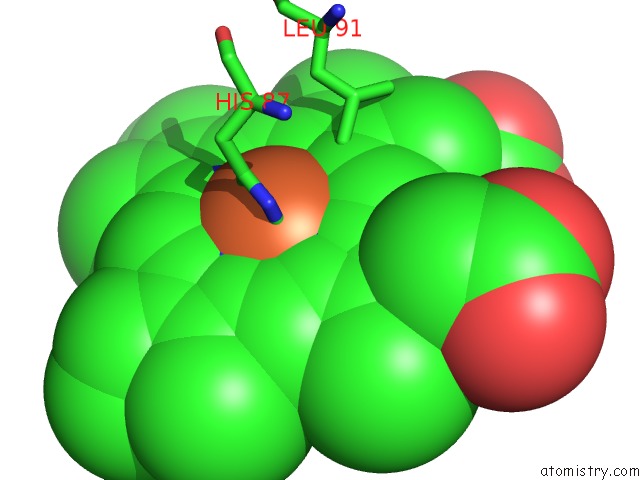
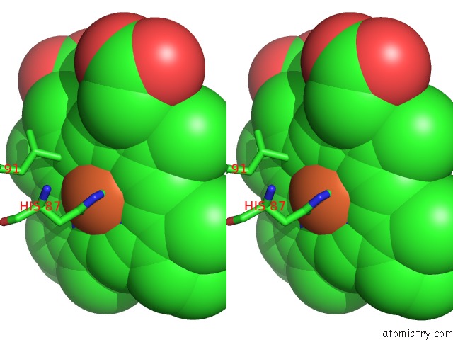
Iron binding site 1 out of 4 in 2w6v
Go back to
Iron binding site 1 out
of 4 in the Structure of Human Deoxy Hemoglobin A in Complex with Xenon
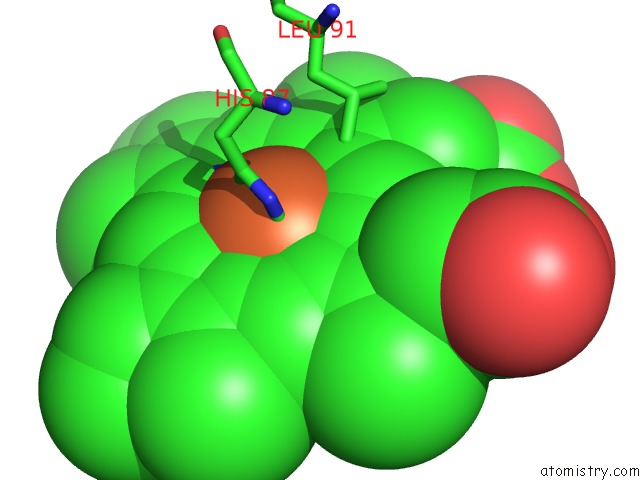
Mono view
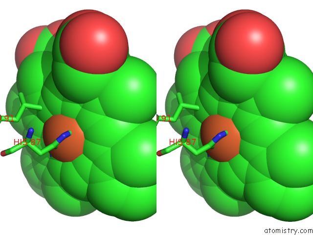
Stereo pair view
Mono view
Stereo pair view
A full contact list of Iron with other atoms in the Fe binding
site number 1 of Structure of Human Deoxy Hemoglobin A in Complex with Xenon within 5.0Å range:
|
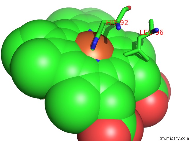
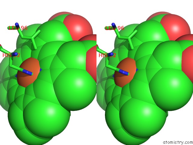
Iron binding site 2 out of 4 in 2w6v
Go back to
Iron binding site 2 out
of 4 in the Structure of Human Deoxy Hemoglobin A in Complex with Xenon
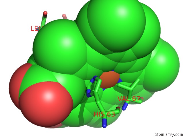
Mono view
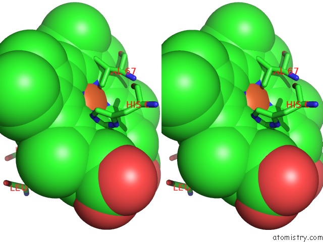
Stereo pair view
Mono view
Stereo pair view
A full contact list of Iron with other atoms in the Fe binding
site number 2 of Structure of Human Deoxy Hemoglobin A in Complex with Xenon within 5.0Å range:
|
Iron binding site 3 out of 4 in 2w6v
Go back to
Iron binding site 3 out
of 4 in the Structure of Human Deoxy Hemoglobin A in Complex with Xenon
Mono view
Stereo pair view
Mono view
Stereo pair view
A full contact list of Iron with other atoms in the Fe binding
site number 3 of Structure of Human Deoxy Hemoglobin A in Complex with Xenon within 5.0Å range:
|
Iron binding site 4 out of 4 in 2w6v
Go back to
Iron binding site 4 out
of 4 in the Structure of Human Deoxy Hemoglobin A in Complex with Xenon
Mono view
Stereo pair view
Mono view
Stereo pair view
A full contact list of Iron with other atoms in the Fe binding
site number 4 of Structure of Human Deoxy Hemoglobin A in Complex with Xenon within 5.0Å range:
|
Reference:
C.Savino,
A.E.Miele,
F.Draghi,
K.A.Johnson,
G.Sciara,
M.Brunori,
B.Vallone.
Pattern of Cavities in Globins: the Case of Human Hemoglobin. Biopolymers V. 91 1097 2009.
ISSN: ISSN 0006-3525
PubMed: 19365817
DOI: 10.1002/BIP.21201
Page generated: Sun Aug 4 03:38:45 2024
ISSN: ISSN 0006-3525
PubMed: 19365817
DOI: 10.1002/BIP.21201
Last articles
Zn in 9MJ5Zn in 9HNW
Zn in 9G0L
Zn in 9FNE
Zn in 9DZN
Zn in 9E0I
Zn in 9D32
Zn in 9DAK
Zn in 8ZXC
Zn in 8ZUF