Iron »
PDB 2w3h-2wl3 »
2wbo »
Iron in PDB 2wbo: Crystal Structure of Vioc in Complex with L-Arginine
Protein crystallography data
The structure of Crystal Structure of Vioc in Complex with L-Arginine, PDB code: 2wbo
was solved by
V.Helmetag,
S.A.Samel,
M.G.Thomas,
M.A.Marahiel,
L.-O.Essen,
with X-Ray Crystallography technique. A brief refinement statistics is given in the table below:
| Resolution Low / High (Å) | 59.03 / 1.30 |
| Space group | C 1 2 1 |
| Cell size a, b, c (Å), α, β, γ (°) | 80.630, 67.340, 62.420, 90.00, 108.83, 90.00 |
| R / Rfree (%) | 16.1 / 21.2 |
Iron Binding Sites:
The binding sites of Iron atom in the Crystal Structure of Vioc in Complex with L-Arginine
(pdb code 2wbo). This binding sites where shown within
5.0 Angstroms radius around Iron atom.
In total 2 binding sites of Iron where determined in the Crystal Structure of Vioc in Complex with L-Arginine, PDB code: 2wbo:
Jump to Iron binding site number: 1; 2;
In total 2 binding sites of Iron where determined in the Crystal Structure of Vioc in Complex with L-Arginine, PDB code: 2wbo:
Jump to Iron binding site number: 1; 2;
Iron binding site 1 out of 2 in 2wbo
Go back to
Iron binding site 1 out
of 2 in the Crystal Structure of Vioc in Complex with L-Arginine
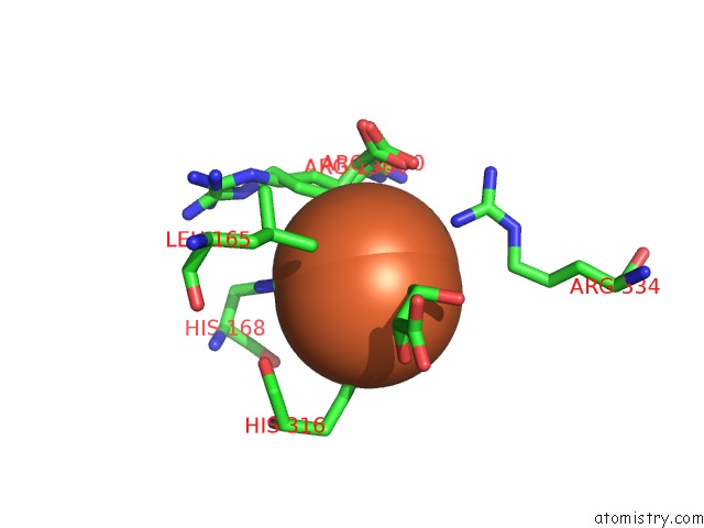
Mono view
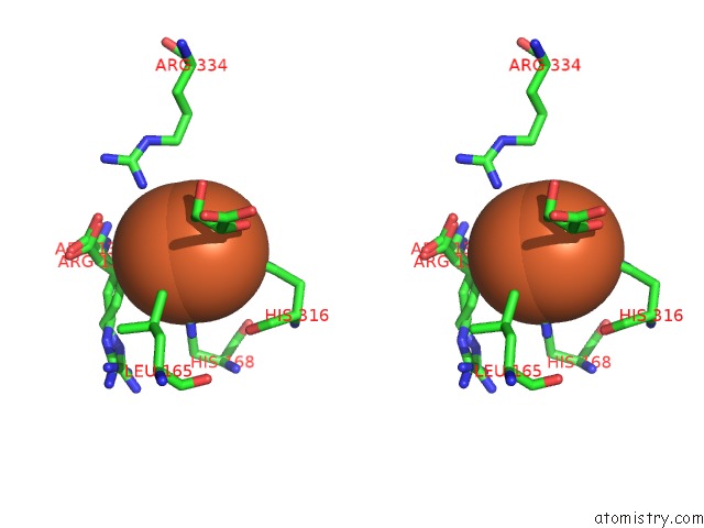
Stereo pair view
Mono view
Stereo pair view
A full contact list of Iron with other atoms in the Fe binding
site number 1 of Crystal Structure of Vioc in Complex with L-Arginine within 5.0Å range:
|
Iron binding site 2 out of 2 in 2wbo
Go back to
Iron binding site 2 out
of 2 in the Crystal Structure of Vioc in Complex with L-Arginine

Mono view

Stereo pair view
Mono view
Stereo pair view
A full contact list of Iron with other atoms in the Fe binding
site number 2 of Crystal Structure of Vioc in Complex with L-Arginine within 5.0Å range:
|
Reference:
V.Helmetag,
S.A.Samel,
M.G.Thomas,
M.A.Marahiel,
L.-O.Essen.
Structural Basis For the Erythro-Stereospecificity of the L-Arginine Oxygenase Vioc in Viomycin Biosynthesis. Febs J. V. 276 3669 2009.
ISSN: ISSN 1742-464X
PubMed: 19490124
DOI: 10.1111/J.1742-4658.2009.07085.X
Page generated: Sun Aug 4 03:43:00 2024
ISSN: ISSN 1742-464X
PubMed: 19490124
DOI: 10.1111/J.1742-4658.2009.07085.X
Last articles
Zn in 9MJ5Zn in 9HNW
Zn in 9G0L
Zn in 9FNE
Zn in 9DZN
Zn in 9E0I
Zn in 9D32
Zn in 9DAK
Zn in 8ZXC
Zn in 8ZUF