Iron »
PDB 2w3h-2wl3 »
2wbp »
Iron in PDB 2wbp: Crystal Structure of Vioc in Complex with Fe(II), (2S,3S)- Hydroxyarginine, and Succinate
Protein crystallography data
The structure of Crystal Structure of Vioc in Complex with Fe(II), (2S,3S)- Hydroxyarginine, and Succinate, PDB code: 2wbp
was solved by
V.Helmetag,
S.A.Samel,
M.G.Thomas,
M.A.Marahiel,
L.-O.Essen,
with X-Ray Crystallography technique. A brief refinement statistics is given in the table below:
| Resolution Low / High (Å) | 19.96 / 1.16 |
| Space group | C 1 2 1 |
| Cell size a, b, c (Å), α, β, γ (°) | 80.770, 66.930, 62.900, 90.00, 109.16, 90.00 |
| R / Rfree (%) | 13.224 / 17.099 |
Iron Binding Sites:
The binding sites of Iron atom in the Crystal Structure of Vioc in Complex with Fe(II), (2S,3S)- Hydroxyarginine, and Succinate
(pdb code 2wbp). This binding sites where shown within
5.0 Angstroms radius around Iron atom.
In total only one binding site of Iron was determined in the Crystal Structure of Vioc in Complex with Fe(II), (2S,3S)- Hydroxyarginine, and Succinate, PDB code: 2wbp:
In total only one binding site of Iron was determined in the Crystal Structure of Vioc in Complex with Fe(II), (2S,3S)- Hydroxyarginine, and Succinate, PDB code: 2wbp:
Iron binding site 1 out of 1 in 2wbp
Go back to
Iron binding site 1 out
of 1 in the Crystal Structure of Vioc in Complex with Fe(II), (2S,3S)- Hydroxyarginine, and Succinate
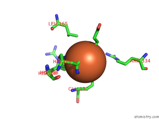
Mono view
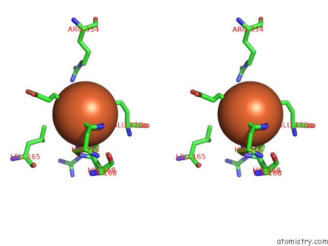
Stereo pair view
Mono view
Stereo pair view
A full contact list of Iron with other atoms in the Fe binding
site number 1 of Crystal Structure of Vioc in Complex with Fe(II), (2S,3S)- Hydroxyarginine, and Succinate within 5.0Å range:
|
Reference:
V.Helmetag,
S.A.Samel,
M.G.Thomas,
M.A.Marahiel,
L.-O.Essen.
Structural Basis For the Erythro-Stereospecificity of the L-Arginine Oxygenase Vioc in Viomycin Biosynthesis. Febs J. V. 276 3669 2009.
ISSN: ISSN 1742-464X
PubMed: 19490124
DOI: 10.1111/J.1742-4658.2009.07085.X
Page generated: Thu Jul 17 04:58:46 2025
ISSN: ISSN 1742-464X
PubMed: 19490124
DOI: 10.1111/J.1742-4658.2009.07085.X
Last articles
Fe in 2YXOFe in 2YRS
Fe in 2YXC
Fe in 2YNM
Fe in 2YVJ
Fe in 2YP1
Fe in 2YU2
Fe in 2YU1
Fe in 2YQB
Fe in 2YOO