Iron »
PDB 2w3h-2wl3 »
2wio »
Iron in PDB 2wio: Structure of the Histidine Tagged, Open Cytochrome P450 Eryk From S. Erythraea
Protein crystallography data
The structure of Structure of the Histidine Tagged, Open Cytochrome P450 Eryk From S. Erythraea, PDB code: 2wio
was solved by
C.Savino,
L.C.Montemiglio,
G.Sciara,
A.E.Miele,
S.G.Kedrew,
S.Gianni,
B.Vallone,
with X-Ray Crystallography technique. A brief refinement statistics is given in the table below:
| Resolution Low / High (Å) | 89.80 / 2.00 |
| Space group | P 21 21 21 |
| Cell size a, b, c (Å), α, β, γ (°) | 37.932, 57.341, 179.557, 90.00, 90.00, 90.00 |
| R / Rfree (%) | 18.341 / 23.202 |
Iron Binding Sites:
The binding sites of Iron atom in the Structure of the Histidine Tagged, Open Cytochrome P450 Eryk From S. Erythraea
(pdb code 2wio). This binding sites where shown within
5.0 Angstroms radius around Iron atom.
In total only one binding site of Iron was determined in the Structure of the Histidine Tagged, Open Cytochrome P450 Eryk From S. Erythraea, PDB code: 2wio:
In total only one binding site of Iron was determined in the Structure of the Histidine Tagged, Open Cytochrome P450 Eryk From S. Erythraea, PDB code: 2wio:
Iron binding site 1 out of 1 in 2wio
Go back to
Iron binding site 1 out
of 1 in the Structure of the Histidine Tagged, Open Cytochrome P450 Eryk From S. Erythraea
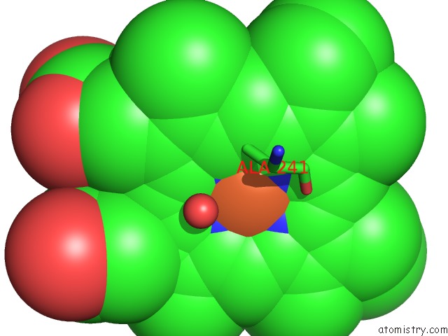
Mono view
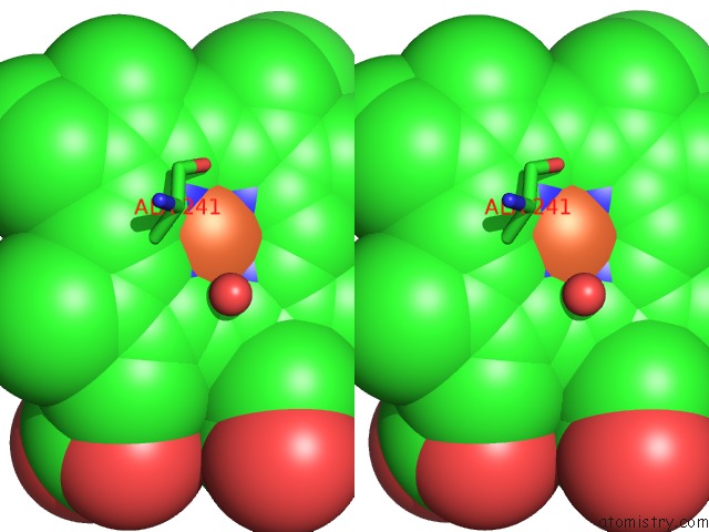
Stereo pair view
Mono view
Stereo pair view
A full contact list of Iron with other atoms in the Fe binding
site number 1 of Structure of the Histidine Tagged, Open Cytochrome P450 Eryk From S. Erythraea within 5.0Å range:
|
Reference:
C.Savino,
L.C.Montemiglio,
G.Sciara,
A.E.Miele,
S.G.Kendrew,
P.Jemth,
S.Gianni,
B.Vallone.
Investigating the Structural Plasticity of A Cytochrome P450: Three-Dimensional Structures of P450 Eryk and Binding to Its Physiological Substrate. J.Biol.Chem. V. 284 29170 2009.
ISSN: ISSN 0021-9258
PubMed: 19625248
DOI: 10.1074/JBC.M109.003590
Page generated: Thu Jul 17 05:03:17 2025
ISSN: ISSN 0021-9258
PubMed: 19625248
DOI: 10.1074/JBC.M109.003590
Last articles
Fe in 2YXOFe in 2YRS
Fe in 2YXC
Fe in 2YNM
Fe in 2YVJ
Fe in 2YP1
Fe in 2YU2
Fe in 2YU1
Fe in 2YQB
Fe in 2YOO