Iron »
PDB 2wl9-2xdq »
2ws3 »
Iron in PDB 2ws3: Crystal Structure of the E. Coli Succinate:Quinone Oxidoreductase (Sqr) Sdhd TYR83PHE Mutant
Enzymatic activity of Crystal Structure of the E. Coli Succinate:Quinone Oxidoreductase (Sqr) Sdhd TYR83PHE Mutant
All present enzymatic activity of Crystal Structure of the E. Coli Succinate:Quinone Oxidoreductase (Sqr) Sdhd TYR83PHE Mutant:
1.3.5.1; 1.3.99.1;
1.3.5.1; 1.3.99.1;
Protein crystallography data
The structure of Crystal Structure of the E. Coli Succinate:Quinone Oxidoreductase (Sqr) Sdhd TYR83PHE Mutant, PDB code: 2ws3
was solved by
J.Ruprecht,
V.Yankovskaya,
E.Maklashina,
S.Iwata,
G.Cecchini,
with X-Ray Crystallography technique. A brief refinement statistics is given in the table below:
| Resolution Low / High (Å) | 49.03 / 3.20 |
| Space group | P 21 21 21 |
| Cell size a, b, c (Å), α, β, γ (°) | 119.850, 184.710, 203.310, 90.00, 90.00, 90.00 |
| R / Rfree (%) | 21.87 / 25.271 |
Other elements in 2ws3:
The structure of Crystal Structure of the E. Coli Succinate:Quinone Oxidoreductase (Sqr) Sdhd TYR83PHE Mutant also contains other interesting chemical elements:
| Sodium | (Na) | 3 atoms |
Iron Binding Sites:
Pages:
>>> Page 1 <<< Page 2, Binding sites: 11 - 20; Page 3, Binding sites: 21 - 30;Binding sites:
The binding sites of Iron atom in the Crystal Structure of the E. Coli Succinate:Quinone Oxidoreductase (Sqr) Sdhd TYR83PHE Mutant (pdb code 2ws3). This binding sites where shown within 5.0 Angstroms radius around Iron atom.In total 30 binding sites of Iron where determined in the Crystal Structure of the E. Coli Succinate:Quinone Oxidoreductase (Sqr) Sdhd TYR83PHE Mutant, PDB code: 2ws3:
Jump to Iron binding site number: 1; 2; 3; 4; 5; 6; 7; 8; 9; 10;
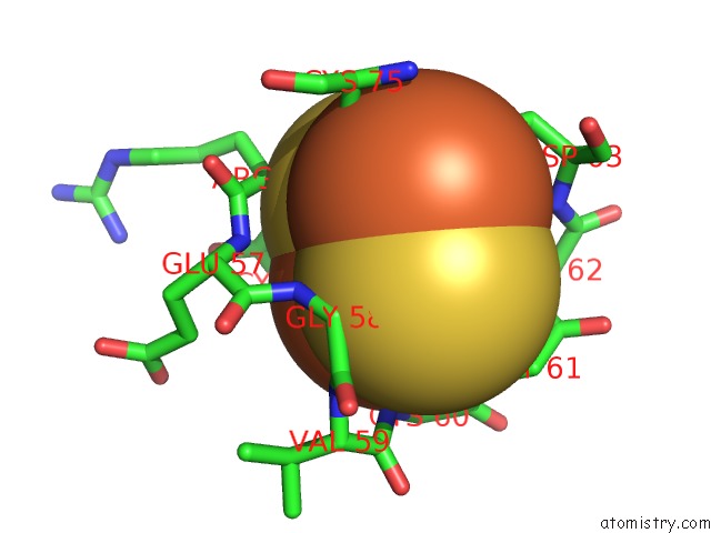
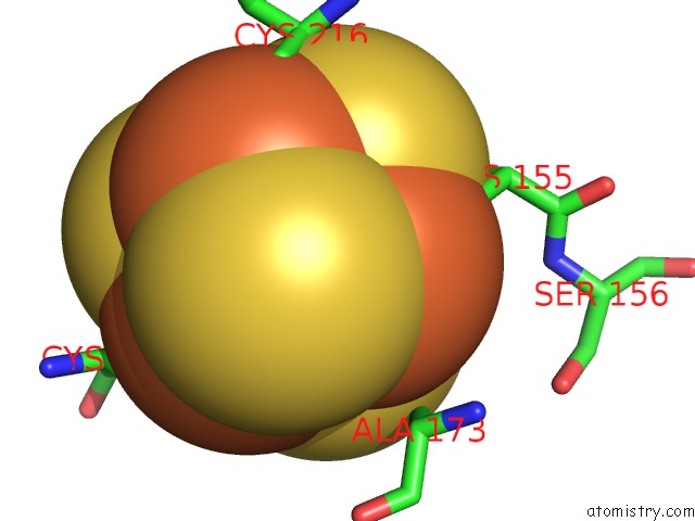
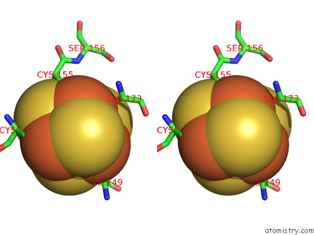
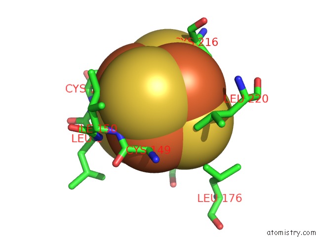
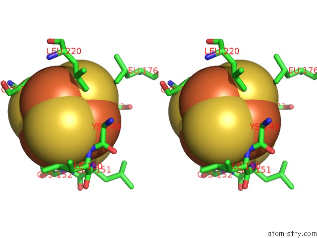
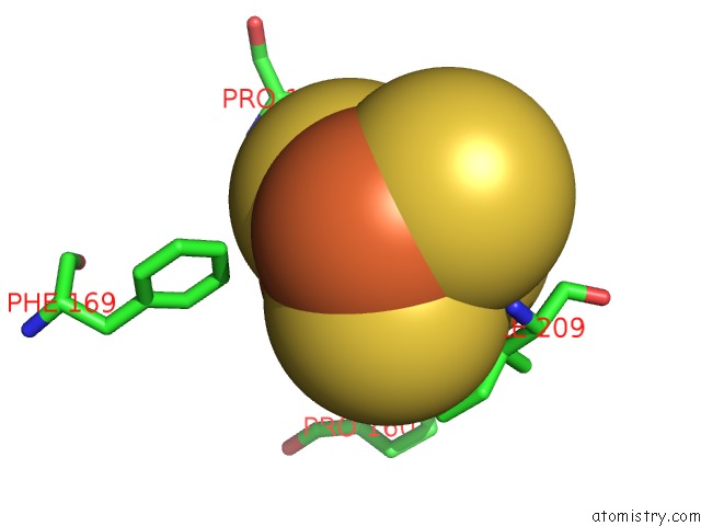
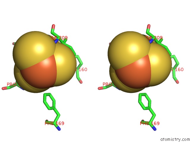
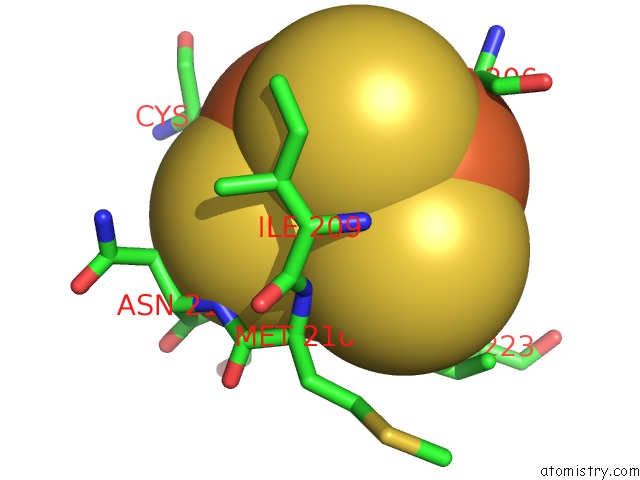
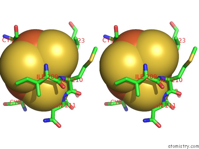
Iron binding site 1 out of 30 in 2ws3
Go back to
Iron binding site 1 out
of 30 in the Crystal Structure of the E. Coli Succinate:Quinone Oxidoreductase (Sqr) Sdhd TYR83PHE Mutant
Mono view
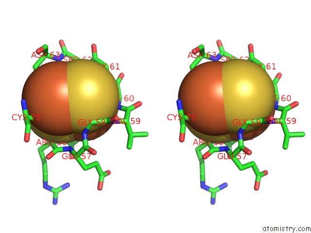
Stereo pair view
Mono view
Stereo pair view
A full contact list of Iron with other atoms in the Fe binding
site number 1 of Crystal Structure of the E. Coli Succinate:Quinone Oxidoreductase (Sqr) Sdhd TYR83PHE Mutant within 5.0Å range:
|
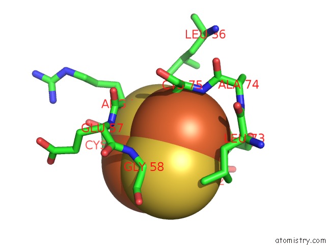
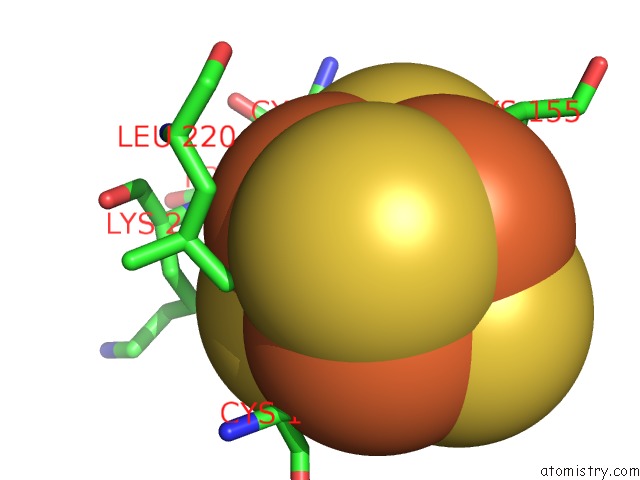
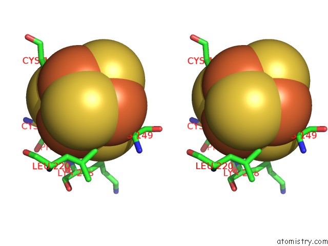
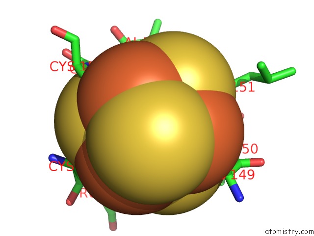
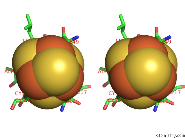
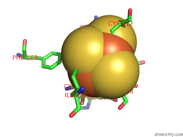
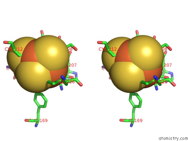
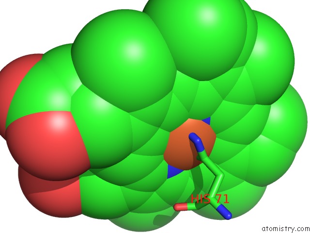
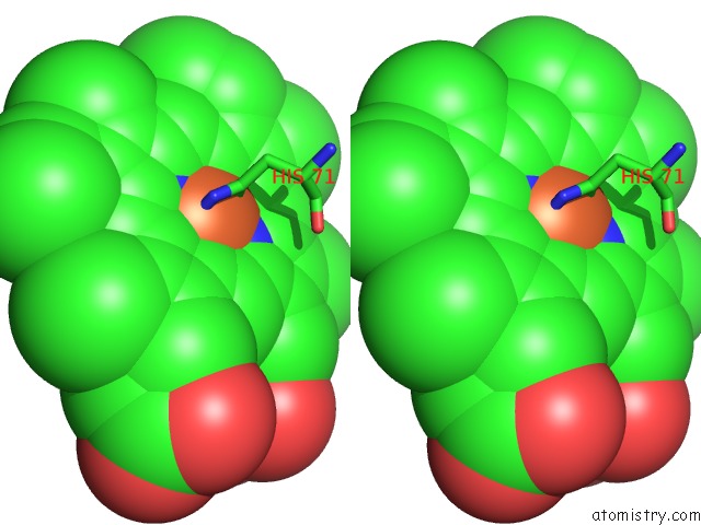
Iron binding site 2 out of 30 in 2ws3
Go back to
Iron binding site 2 out
of 30 in the Crystal Structure of the E. Coli Succinate:Quinone Oxidoreductase (Sqr) Sdhd TYR83PHE Mutant
Mono view
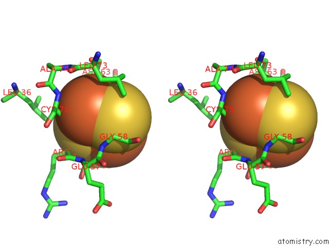
Stereo pair view
Mono view
Stereo pair view
A full contact list of Iron with other atoms in the Fe binding
site number 2 of Crystal Structure of the E. Coli Succinate:Quinone Oxidoreductase (Sqr) Sdhd TYR83PHE Mutant within 5.0Å range:
|
Iron binding site 3 out of 30 in 2ws3
Go back to
Iron binding site 3 out
of 30 in the Crystal Structure of the E. Coli Succinate:Quinone Oxidoreductase (Sqr) Sdhd TYR83PHE Mutant
Mono view
Stereo pair view
Mono view
Stereo pair view
A full contact list of Iron with other atoms in the Fe binding
site number 3 of Crystal Structure of the E. Coli Succinate:Quinone Oxidoreductase (Sqr) Sdhd TYR83PHE Mutant within 5.0Å range:
|
Iron binding site 4 out of 30 in 2ws3
Go back to
Iron binding site 4 out
of 30 in the Crystal Structure of the E. Coli Succinate:Quinone Oxidoreductase (Sqr) Sdhd TYR83PHE Mutant
Mono view
Stereo pair view
Mono view
Stereo pair view
A full contact list of Iron with other atoms in the Fe binding
site number 4 of Crystal Structure of the E. Coli Succinate:Quinone Oxidoreductase (Sqr) Sdhd TYR83PHE Mutant within 5.0Å range:
|
Iron binding site 5 out of 30 in 2ws3
Go back to
Iron binding site 5 out
of 30 in the Crystal Structure of the E. Coli Succinate:Quinone Oxidoreductase (Sqr) Sdhd TYR83PHE Mutant
Mono view
Stereo pair view
Mono view
Stereo pair view
A full contact list of Iron with other atoms in the Fe binding
site number 5 of Crystal Structure of the E. Coli Succinate:Quinone Oxidoreductase (Sqr) Sdhd TYR83PHE Mutant within 5.0Å range:
|
Iron binding site 6 out of 30 in 2ws3
Go back to
Iron binding site 6 out
of 30 in the Crystal Structure of the E. Coli Succinate:Quinone Oxidoreductase (Sqr) Sdhd TYR83PHE Mutant
Mono view
Stereo pair view
Mono view
Stereo pair view
A full contact list of Iron with other atoms in the Fe binding
site number 6 of Crystal Structure of the E. Coli Succinate:Quinone Oxidoreductase (Sqr) Sdhd TYR83PHE Mutant within 5.0Å range:
|
Iron binding site 7 out of 30 in 2ws3
Go back to
Iron binding site 7 out
of 30 in the Crystal Structure of the E. Coli Succinate:Quinone Oxidoreductase (Sqr) Sdhd TYR83PHE Mutant
Mono view
Stereo pair view
Mono view
Stereo pair view
A full contact list of Iron with other atoms in the Fe binding
site number 7 of Crystal Structure of the E. Coli Succinate:Quinone Oxidoreductase (Sqr) Sdhd TYR83PHE Mutant within 5.0Å range:
|
Iron binding site 8 out of 30 in 2ws3
Go back to
Iron binding site 8 out
of 30 in the Crystal Structure of the E. Coli Succinate:Quinone Oxidoreductase (Sqr) Sdhd TYR83PHE Mutant
Mono view
Stereo pair view
Mono view
Stereo pair view
A full contact list of Iron with other atoms in the Fe binding
site number 8 of Crystal Structure of the E. Coli Succinate:Quinone Oxidoreductase (Sqr) Sdhd TYR83PHE Mutant within 5.0Å range:
|
Iron binding site 9 out of 30 in 2ws3
Go back to
Iron binding site 9 out
of 30 in the Crystal Structure of the E. Coli Succinate:Quinone Oxidoreductase (Sqr) Sdhd TYR83PHE Mutant
Mono view
Stereo pair view
Mono view
Stereo pair view
A full contact list of Iron with other atoms in the Fe binding
site number 9 of Crystal Structure of the E. Coli Succinate:Quinone Oxidoreductase (Sqr) Sdhd TYR83PHE Mutant within 5.0Å range:
|
Iron binding site 10 out of 30 in 2ws3
Go back to
Iron binding site 10 out
of 30 in the Crystal Structure of the E. Coli Succinate:Quinone Oxidoreductase (Sqr) Sdhd TYR83PHE Mutant
Mono view
Stereo pair view
Mono view
Stereo pair view
A full contact list of Iron with other atoms in the Fe binding
site number 10 of Crystal Structure of the E. Coli Succinate:Quinone Oxidoreductase (Sqr) Sdhd TYR83PHE Mutant within 5.0Å range:
|
Reference:
J.Ruprecht,
V.Yankovskaya,
E.Maklashina,
S.Iwata,
G.Cecchini.
Succinate Dehydrogenase Activity To Be Published.
Page generated: Sun Aug 4 04:04:46 2024
Last articles
Zn in 9MJ5Zn in 9HNW
Zn in 9G0L
Zn in 9FNE
Zn in 9DZN
Zn in 9E0I
Zn in 9D32
Zn in 9DAK
Zn in 8ZXC
Zn in 8ZUF