Iron »
PDB 2xf2-2xuz »
2xl8 »
Iron in PDB 2xl8: Reduced Structure of R124F Mutant of Cytochrome C' From Alcaligenes Xylosoxidans
Protein crystallography data
The structure of Reduced Structure of R124F Mutant of Cytochrome C' From Alcaligenes Xylosoxidans, PDB code: 2xl8
was solved by
M.A.Hough,
S.V.Antonyuk,
S.Barbieri,
N.Rustage,
A.L.Mckay,
A.E.Servid,
R.R.Eady,
C.R.Andrew,
S.S.Hasnain,
with X-Ray Crystallography technique. A brief refinement statistics is given in the table below:
| Resolution Low / High (Å) | 46.47 / 1.14 |
| Space group | P 65 2 2 |
| Cell size a, b, c (Å), α, β, γ (°) | 53.681, 53.681, 181.205, 90.00, 90.00, 120.00 |
| R / Rfree (%) | 14 / 16.1 |
Iron Binding Sites:
The binding sites of Iron atom in the Reduced Structure of R124F Mutant of Cytochrome C' From Alcaligenes Xylosoxidans
(pdb code 2xl8). This binding sites where shown within
5.0 Angstroms radius around Iron atom.
In total only one binding site of Iron was determined in the Reduced Structure of R124F Mutant of Cytochrome C' From Alcaligenes Xylosoxidans, PDB code: 2xl8:
In total only one binding site of Iron was determined in the Reduced Structure of R124F Mutant of Cytochrome C' From Alcaligenes Xylosoxidans, PDB code: 2xl8:
Iron binding site 1 out of 1 in 2xl8
Go back to
Iron binding site 1 out
of 1 in the Reduced Structure of R124F Mutant of Cytochrome C' From Alcaligenes Xylosoxidans
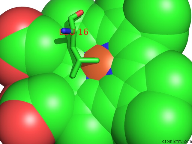
Mono view
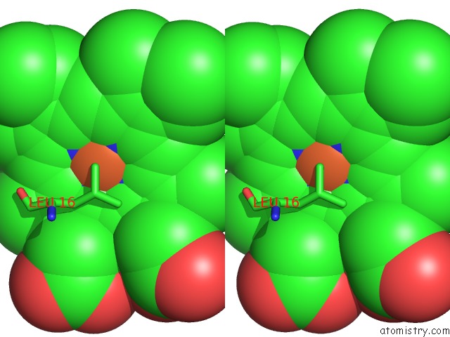
Stereo pair view
Mono view
Stereo pair view
A full contact list of Iron with other atoms in the Fe binding
site number 1 of Reduced Structure of R124F Mutant of Cytochrome C' From Alcaligenes Xylosoxidans within 5.0Å range:
|
Reference:
M.A.Hough,
S.V.Antonyuk,
S.Barbieri,
N.Rustage,
A.L.Mckay,
A.E.Servid,
R.R.Eady,
C.R.Andrew,
S.S.Hasnain.
Distal-to-Proximal No Conversion in Hemoproteins: the Role of the Proximal Pocket. J.Mol.Biol. V. 405 395 2011.
ISSN: ISSN 0022-2836
PubMed: 21073879
DOI: 10.1016/J.JMB.2010.10.035
Page generated: Sun Aug 4 04:38:09 2024
ISSN: ISSN 0022-2836
PubMed: 21073879
DOI: 10.1016/J.JMB.2010.10.035
Last articles
Zn in 9J0NZn in 9J0O
Zn in 9J0P
Zn in 9FJX
Zn in 9EKB
Zn in 9C0F
Zn in 9CAH
Zn in 9CH0
Zn in 9CH3
Zn in 9CH1