Iron »
PDB 2xf2-2xuz »
2xue »
Iron in PDB 2xue: Crystal Structure of JMJD3
Protein crystallography data
The structure of Crystal Structure of JMJD3, PDB code: 2xue
was solved by
C.Chung,
P.Rowland,
J.Mosley,
P.J.Thomas,
with X-Ray Crystallography technique. A brief refinement statistics is given in the table below:
| Resolution Low / High (Å) | 71.13 / 2.00 |
| Space group | P 1 |
| Cell size a, b, c (Å), α, β, γ (°) | 61.215, 65.154, 77.462, 86.09, 67.19, 68.26 |
| R / Rfree (%) | 17.932 / 21.574 |
Other elements in 2xue:
The structure of Crystal Structure of JMJD3 also contains other interesting chemical elements:
| Zinc | (Zn) | 2 atoms |
Iron Binding Sites:
The binding sites of Iron atom in the Crystal Structure of JMJD3
(pdb code 2xue). This binding sites where shown within
5.0 Angstroms radius around Iron atom.
In total 2 binding sites of Iron where determined in the Crystal Structure of JMJD3, PDB code: 2xue:
Jump to Iron binding site number: 1; 2;
In total 2 binding sites of Iron where determined in the Crystal Structure of JMJD3, PDB code: 2xue:
Jump to Iron binding site number: 1; 2;
Iron binding site 1 out of 2 in 2xue
Go back to
Iron binding site 1 out
of 2 in the Crystal Structure of JMJD3
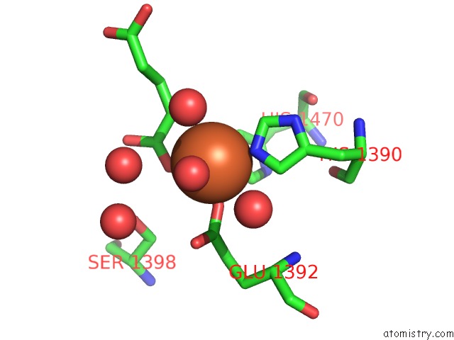
Mono view
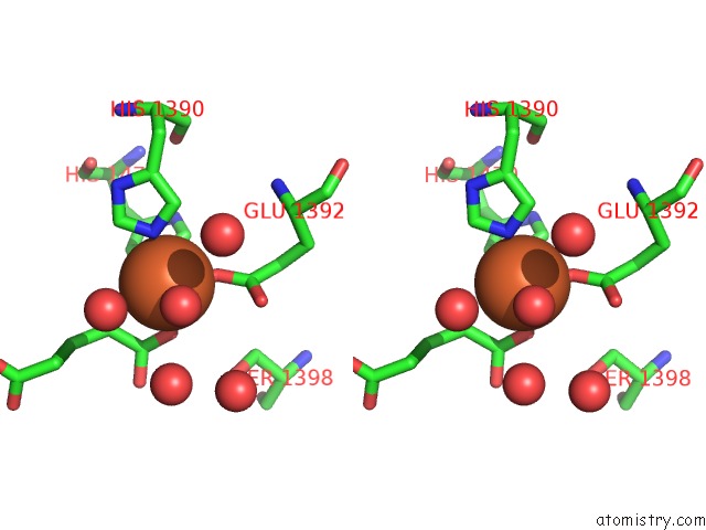
Stereo pair view
Mono view
Stereo pair view
A full contact list of Iron with other atoms in the Fe binding
site number 1 of Crystal Structure of JMJD3 within 5.0Å range:
|
Iron binding site 2 out of 2 in 2xue
Go back to
Iron binding site 2 out
of 2 in the Crystal Structure of JMJD3
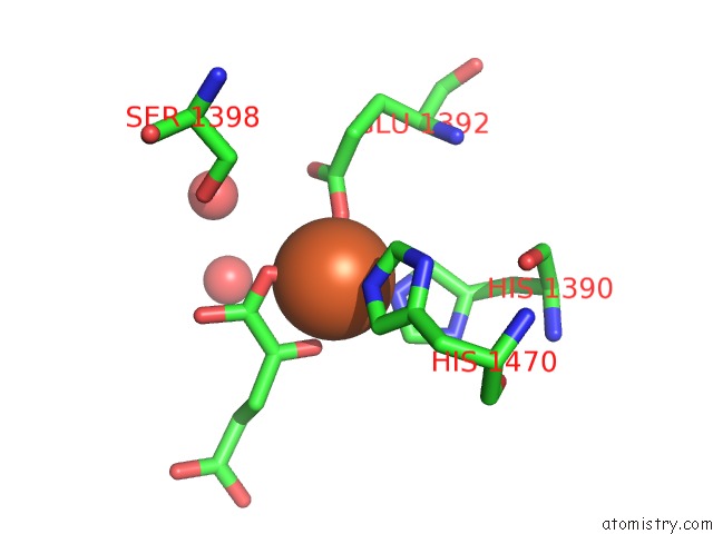
Mono view
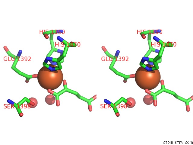
Stereo pair view
Mono view
Stereo pair view
A full contact list of Iron with other atoms in the Fe binding
site number 2 of Crystal Structure of JMJD3 within 5.0Å range:
|
Reference:
L.Kruidenier,
C.Chung,
Z.Cheng,
J.Liddle,
K.Che,
G.Joberty,
M.Bantscheff,
C.Bountra,
A.Bridges,
H.Diallo,
D.Eberhard,
S.Hutchinson,
E.Jones,
R.Katso,
M.Leveridge,
P.K.Mander,
J.Mosley,
C.Ramirez-Molina,
P.Rowland,
C.J.Schofield,
R.J.Sheppard,
J.E.Smith,
C.Swales,
R.Tanner,
P.Thomas,
A.Tumber,
G.Drewes,
U.Oppermann,
D.J.Patel,
K.Lee,
D.M.Wilson.
A Selective Jumonji H3K27 Demethylase Inhibitor Modulates the Proinflammatory Macrophage Response Nature V. 488 404 2012.
ISSN: ISSN 0028-0836
PubMed: 22842901
DOI: 10.1038/NATURE11262
Page generated: Sun Aug 4 04:43:12 2024
ISSN: ISSN 0028-0836
PubMed: 22842901
DOI: 10.1038/NATURE11262
Last articles
Zn in 9MJ5Zn in 9HNW
Zn in 9G0L
Zn in 9FNE
Zn in 9DZN
Zn in 9E0I
Zn in 9D32
Zn in 9DAK
Zn in 8ZXC
Zn in 8ZUF