Iron »
PDB 2xv1-2yde »
2y5a »
Iron in PDB 2y5a: Cytochrome C Peroxidase (Ccp) W191G Bound to 3-Aminopyridine
Enzymatic activity of Cytochrome C Peroxidase (Ccp) W191G Bound to 3-Aminopyridine
All present enzymatic activity of Cytochrome C Peroxidase (Ccp) W191G Bound to 3-Aminopyridine:
1.11.1.5;
1.11.1.5;
Protein crystallography data
The structure of Cytochrome C Peroxidase (Ccp) W191G Bound to 3-Aminopyridine, PDB code: 2y5a
was solved by
D.Cappel,
R.Wahlstrom,
R.Brenk,
C.A.Sotriffer,
with X-Ray Crystallography technique. A brief refinement statistics is given in the table below:
| Resolution Low / High (Å) | 25.18 / 1.25 |
| Space group | P 21 21 21 |
| Cell size a, b, c (Å), α, β, γ (°) | 50.853, 75.362, 106.884, 90.00, 90.00, 90.00 |
| R / Rfree (%) | 14.952 / 16.388 |
Iron Binding Sites:
The binding sites of Iron atom in the Cytochrome C Peroxidase (Ccp) W191G Bound to 3-Aminopyridine
(pdb code 2y5a). This binding sites where shown within
5.0 Angstroms radius around Iron atom.
In total only one binding site of Iron was determined in the Cytochrome C Peroxidase (Ccp) W191G Bound to 3-Aminopyridine, PDB code: 2y5a:
In total only one binding site of Iron was determined in the Cytochrome C Peroxidase (Ccp) W191G Bound to 3-Aminopyridine, PDB code: 2y5a:
Iron binding site 1 out of 1 in 2y5a
Go back to
Iron binding site 1 out
of 1 in the Cytochrome C Peroxidase (Ccp) W191G Bound to 3-Aminopyridine
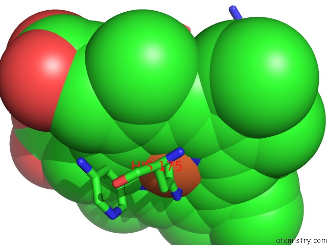
Mono view
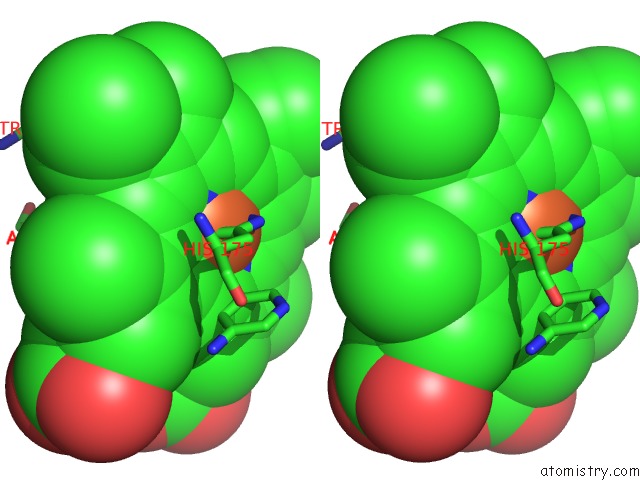
Stereo pair view
Mono view
Stereo pair view
A full contact list of Iron with other atoms in the Fe binding
site number 1 of Cytochrome C Peroxidase (Ccp) W191G Bound to 3-Aminopyridine within 5.0Å range:
|
Reference:
D.Cappel,
R.Wahlstrom,
R.Brenk,
C.A.Sotriffer.
Probing the Dynamic Nature of Water Molecules and Their Influences on Ligand Binding in A Model Binding Site. J.Chem.Inf.Model V. 51 2581 2011.
ISSN: ISSN 1549-9596
PubMed: 21916516
DOI: 10.1021/CI200052J
Page generated: Sun Aug 4 05:05:35 2024
ISSN: ISSN 1549-9596
PubMed: 21916516
DOI: 10.1021/CI200052J
Last articles
F in 7NTHF in 7NTI
F in 7NPC
F in 7NRG
F in 7NR5
F in 7NQS
F in 7NOS
F in 7NP5
F in 7NDV
F in 7NP6