Iron »
PDB 2yeq-2z4g »
2yfi »
Iron in PDB 2yfi: Crystal Structure of Biphenyl Dioxygenase Variant RR41 (Bpdo-RR41)
Enzymatic activity of Crystal Structure of Biphenyl Dioxygenase Variant RR41 (Bpdo-RR41)
All present enzymatic activity of Crystal Structure of Biphenyl Dioxygenase Variant RR41 (Bpdo-RR41):
1.14.12.18;
1.14.12.18;
Protein crystallography data
The structure of Crystal Structure of Biphenyl Dioxygenase Variant RR41 (Bpdo-RR41), PDB code: 2yfi
was solved by
P.Kumar,
J.T.Bolin,
with X-Ray Crystallography technique. A brief refinement statistics is given in the table below:
| Resolution Low / High (Å) | 138.68 / 2.15 |
| Space group | P 1 21 1 |
| Cell size a, b, c (Å), α, β, γ (°) | 86.969, 277.808, 92.933, 90.00, 117.61, 90.00 |
| R / Rfree (%) | 19.992 / 23.163 |
Iron Binding Sites:
Pages:
>>> Page 1 <<< Page 2, Binding sites: 11 - 18;Binding sites:
The binding sites of Iron atom in the Crystal Structure of Biphenyl Dioxygenase Variant RR41 (Bpdo-RR41) (pdb code 2yfi). This binding sites where shown within 5.0 Angstroms radius around Iron atom.In total 18 binding sites of Iron where determined in the Crystal Structure of Biphenyl Dioxygenase Variant RR41 (Bpdo-RR41), PDB code: 2yfi:
Jump to Iron binding site number: 1; 2; 3; 4; 5; 6; 7; 8; 9; 10;
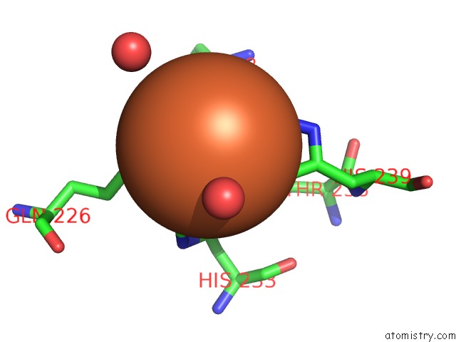
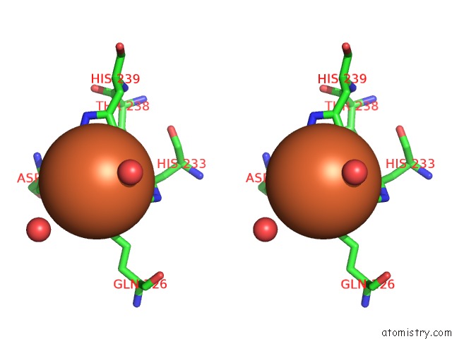
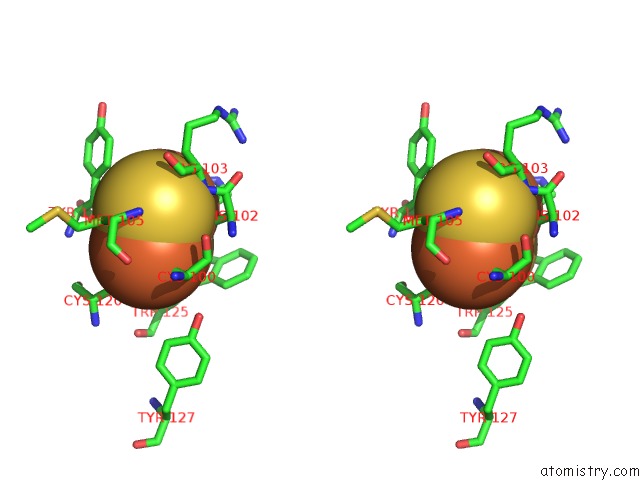
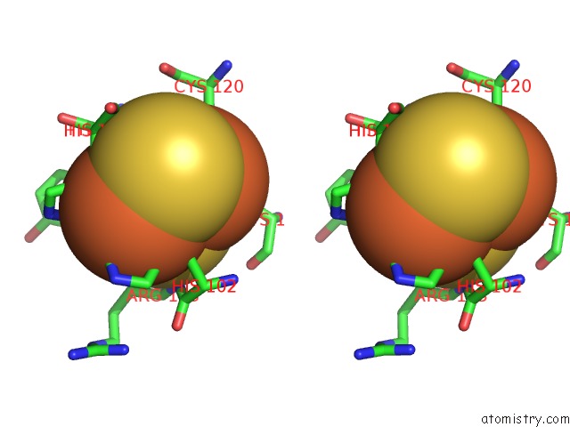
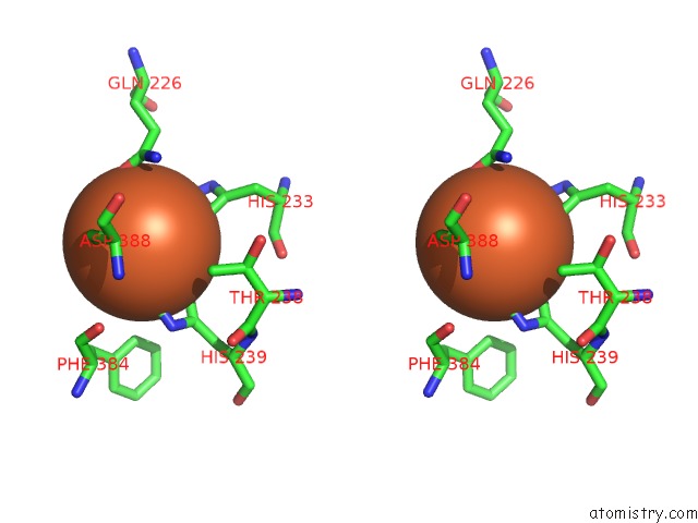
Iron binding site 1 out of 18 in 2yfi
Go back to
Iron binding site 1 out
of 18 in the Crystal Structure of Biphenyl Dioxygenase Variant RR41 (Bpdo-RR41)
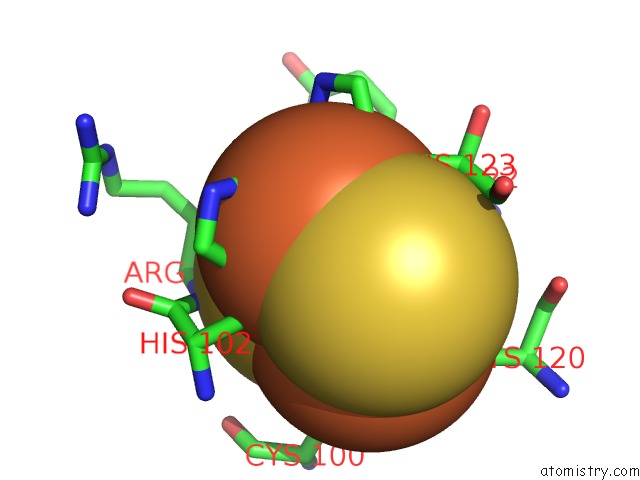
Mono view
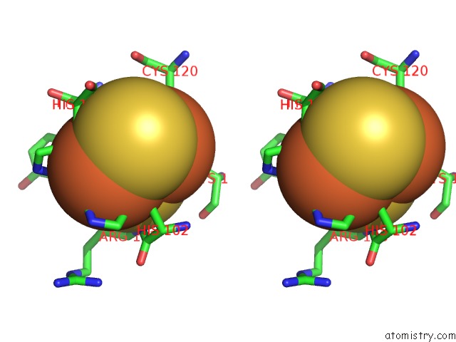
Stereo pair view
Mono view
Stereo pair view
A full contact list of Iron with other atoms in the Fe binding
site number 1 of Crystal Structure of Biphenyl Dioxygenase Variant RR41 (Bpdo-RR41) within 5.0Å range:
|
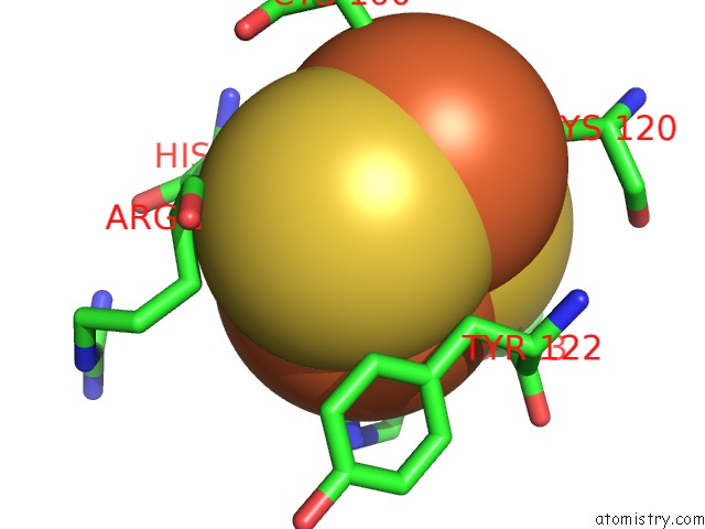
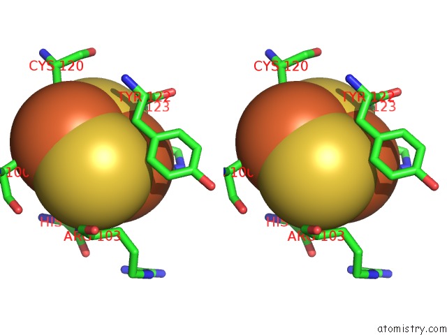
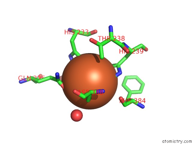
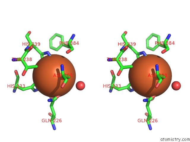
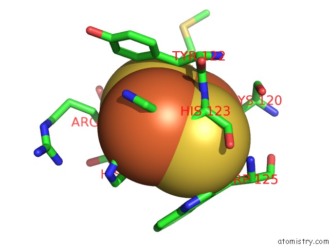
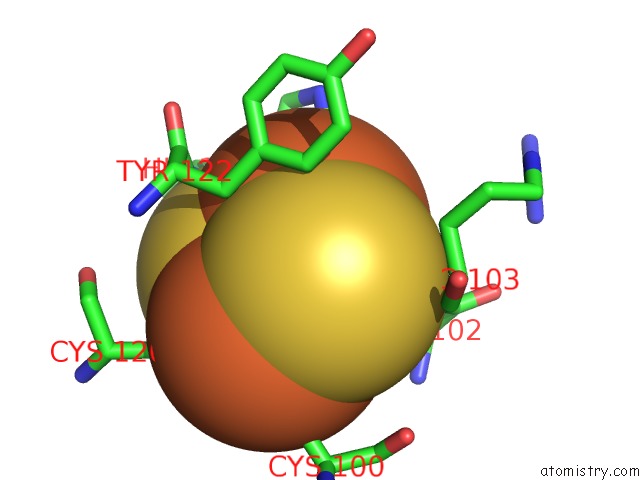
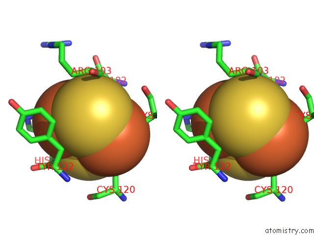
Iron binding site 2 out of 18 in 2yfi
Go back to
Iron binding site 2 out
of 18 in the Crystal Structure of Biphenyl Dioxygenase Variant RR41 (Bpdo-RR41)
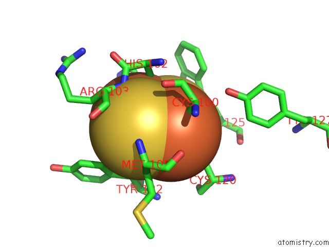
Mono view
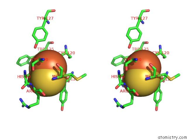
Stereo pair view
Mono view
Stereo pair view
A full contact list of Iron with other atoms in the Fe binding
site number 2 of Crystal Structure of Biphenyl Dioxygenase Variant RR41 (Bpdo-RR41) within 5.0Å range:
|
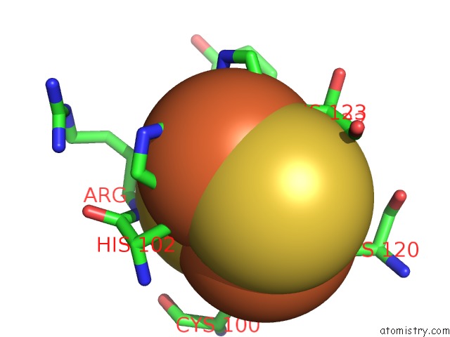
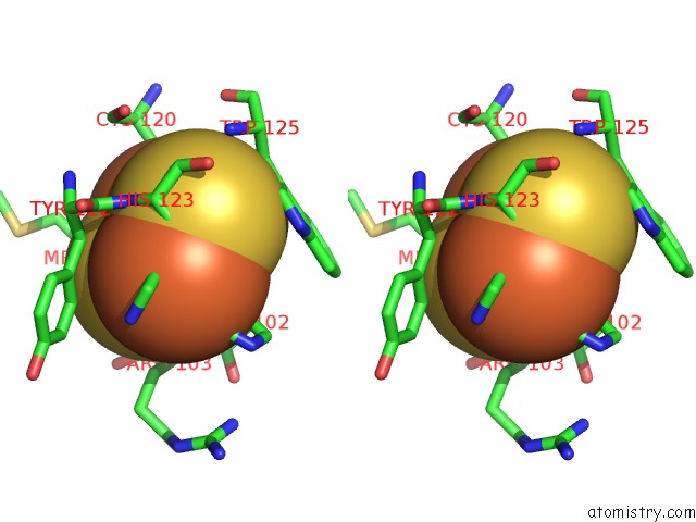
Iron binding site 3 out of 18 in 2yfi
Go back to
Iron binding site 3 out
of 18 in the Crystal Structure of Biphenyl Dioxygenase Variant RR41 (Bpdo-RR41)
Mono view
Stereo pair view
Mono view
Stereo pair view
A full contact list of Iron with other atoms in the Fe binding
site number 3 of Crystal Structure of Biphenyl Dioxygenase Variant RR41 (Bpdo-RR41) within 5.0Å range:
|
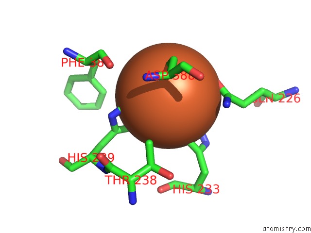
Iron binding site 4 out of 18 in 2yfi
Go back to
Iron binding site 4 out
of 18 in the Crystal Structure of Biphenyl Dioxygenase Variant RR41 (Bpdo-RR41)
Mono view
Stereo pair view
Mono view
Stereo pair view
A full contact list of Iron with other atoms in the Fe binding
site number 4 of Crystal Structure of Biphenyl Dioxygenase Variant RR41 (Bpdo-RR41) within 5.0Å range:
|
Iron binding site 5 out of 18 in 2yfi
Go back to
Iron binding site 5 out
of 18 in the Crystal Structure of Biphenyl Dioxygenase Variant RR41 (Bpdo-RR41)

Mono view
Stereo pair view
Mono view
Stereo pair view
A full contact list of Iron with other atoms in the Fe binding
site number 5 of Crystal Structure of Biphenyl Dioxygenase Variant RR41 (Bpdo-RR41) within 5.0Å range:
|
Iron binding site 6 out of 18 in 2yfi
Go back to
Iron binding site 6 out
of 18 in the Crystal Structure of Biphenyl Dioxygenase Variant RR41 (Bpdo-RR41)
Mono view
Stereo pair view
Mono view
Stereo pair view
A full contact list of Iron with other atoms in the Fe binding
site number 6 of Crystal Structure of Biphenyl Dioxygenase Variant RR41 (Bpdo-RR41) within 5.0Å range:
|
Iron binding site 7 out of 18 in 2yfi
Go back to
Iron binding site 7 out
of 18 in the Crystal Structure of Biphenyl Dioxygenase Variant RR41 (Bpdo-RR41)
Mono view
Stereo pair view
Mono view
Stereo pair view
A full contact list of Iron with other atoms in the Fe binding
site number 7 of Crystal Structure of Biphenyl Dioxygenase Variant RR41 (Bpdo-RR41) within 5.0Å range:
|
Iron binding site 8 out of 18 in 2yfi
Go back to
Iron binding site 8 out
of 18 in the Crystal Structure of Biphenyl Dioxygenase Variant RR41 (Bpdo-RR41)
Mono view
Stereo pair view
Mono view
Stereo pair view
A full contact list of Iron with other atoms in the Fe binding
site number 8 of Crystal Structure of Biphenyl Dioxygenase Variant RR41 (Bpdo-RR41) within 5.0Å range:
|
Iron binding site 9 out of 18 in 2yfi
Go back to
Iron binding site 9 out
of 18 in the Crystal Structure of Biphenyl Dioxygenase Variant RR41 (Bpdo-RR41)
Mono view
Stereo pair view
Mono view
Stereo pair view
A full contact list of Iron with other atoms in the Fe binding
site number 9 of Crystal Structure of Biphenyl Dioxygenase Variant RR41 (Bpdo-RR41) within 5.0Å range:
|
Iron binding site 10 out of 18 in 2yfi
Go back to
Iron binding site 10 out
of 18 in the Crystal Structure of Biphenyl Dioxygenase Variant RR41 (Bpdo-RR41)
Mono view
Stereo pair view
Mono view
Stereo pair view
A full contact list of Iron with other atoms in the Fe binding
site number 10 of Crystal Structure of Biphenyl Dioxygenase Variant RR41 (Bpdo-RR41) within 5.0Å range:
|
Reference:
M.Mohammadi,
J.Viger,
P.Kumar,
D.Barriault,
J.T.Bolin,
M.Sylvestre.
Retuning Rieske-Type Oxygenases to Expand Substrate Range. J.Biol.Chem. V. 286 27612 2011.
ISSN: ISSN 0021-9258
PubMed: 21653696
DOI: 10.1074/JBC.M111.255174
Page generated: Thu Jul 17 06:05:12 2025
ISSN: ISSN 0021-9258
PubMed: 21653696
DOI: 10.1074/JBC.M111.255174
Last articles
Fe in 2YXOFe in 2YRS
Fe in 2YXC
Fe in 2YNM
Fe in 2YVJ
Fe in 2YP1
Fe in 2YU2
Fe in 2YU1
Fe in 2YQB
Fe in 2YOO