Iron »
PDB 2yeq-2z4g »
2yk3 »
Iron in PDB 2yk3: Crithidia Fasciculata Cytochrome C
Protein crystallography data
The structure of Crithidia Fasciculata Cytochrome C, PDB code: 2yk3
was solved by
V.Fulop,
K.A.Sam,
S.J.Ferguson,
M.L.Ginger,
J.W.A.Allen,
with X-Ray Crystallography technique. A brief refinement statistics is given in the table below:
| Resolution Low / High (Å) | 55.81 / 1.55 |
| Space group | C 1 2 1 |
| Cell size a, b, c (Å), α, β, γ (°) | 85.930, 110.880, 60.930, 90.00, 131.20, 90.00 |
| R / Rfree (%) | 20.6 / 24.1 |
Iron Binding Sites:
The binding sites of Iron atom in the Crithidia Fasciculata Cytochrome C
(pdb code 2yk3). This binding sites where shown within
5.0 Angstroms radius around Iron atom.
In total 3 binding sites of Iron where determined in the Crithidia Fasciculata Cytochrome C, PDB code: 2yk3:
Jump to Iron binding site number: 1; 2; 3;
In total 3 binding sites of Iron where determined in the Crithidia Fasciculata Cytochrome C, PDB code: 2yk3:
Jump to Iron binding site number: 1; 2; 3;
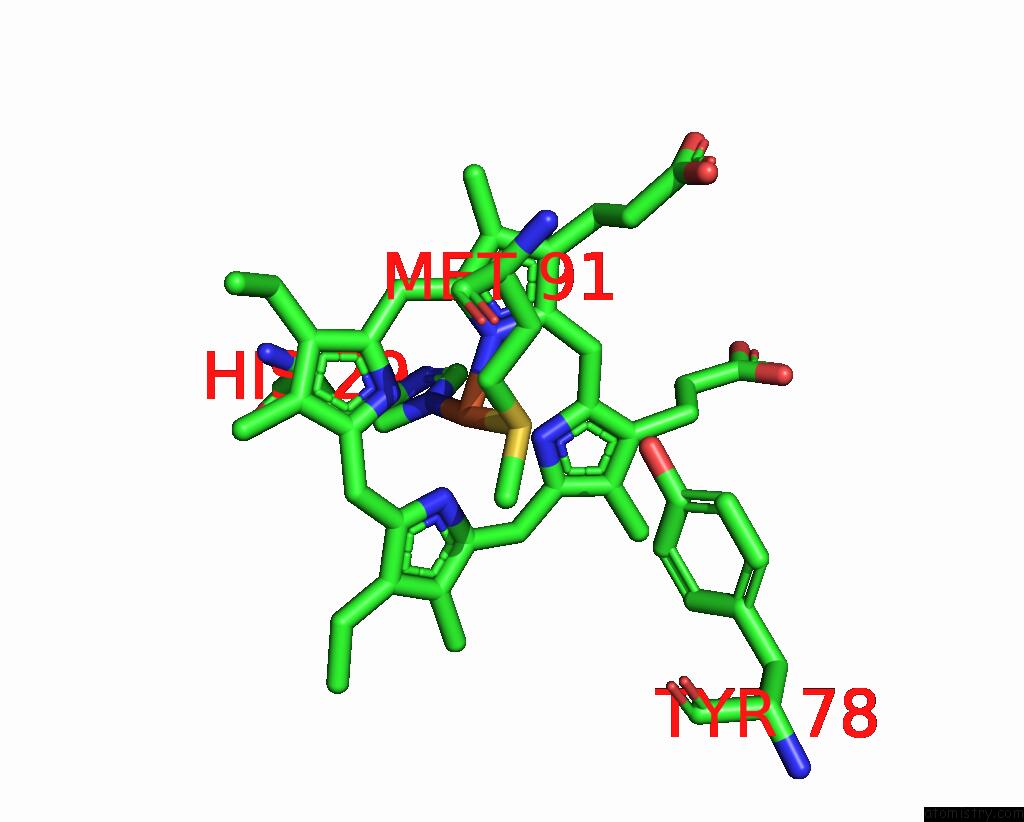
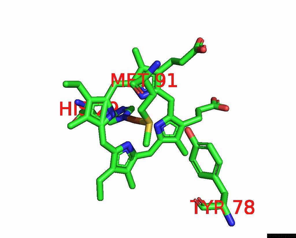
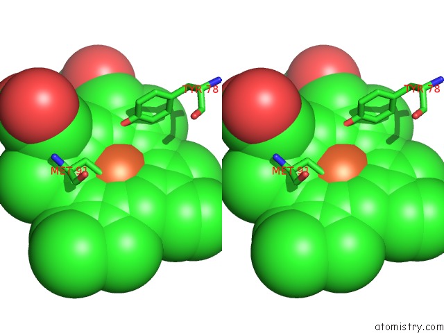
Iron binding site 1 out of 3 in 2yk3
Go back to
Iron binding site 1 out
of 3 in the Crithidia Fasciculata Cytochrome C
Mono view
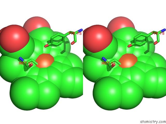
Stereo pair view
Mono view
Stereo pair view
A full contact list of Iron with other atoms in the Fe binding
site number 1 of Crithidia Fasciculata Cytochrome C within 5.0Å range:
|
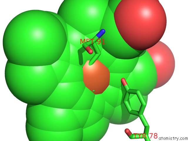
Iron binding site 2 out of 3 in 2yk3
Go back to
Iron binding site 2 out
of 3 in the Crithidia Fasciculata Cytochrome C
Mono view
Stereo pair view
Mono view
Stereo pair view
A full contact list of Iron with other atoms in the Fe binding
site number 2 of Crithidia Fasciculata Cytochrome C within 5.0Å range:
|
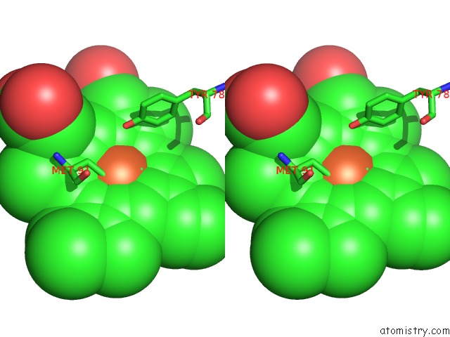
Iron binding site 3 out of 3 in 2yk3
Go back to
Iron binding site 3 out
of 3 in the Crithidia Fasciculata Cytochrome C
Mono view
Stereo pair view
Mono view
Stereo pair view
A full contact list of Iron with other atoms in the Fe binding
site number 3 of Crithidia Fasciculata Cytochrome C within 5.0Å range:
|
Reference:
V.Fulop,
K.A.Sam,
S.J.Ferguson,
M.L.Ginger,
J.W.A.Allen.
Structure of A Trypanosomatid Mitochondrial Cytochrome C with Heme Attached Via Only One Thioether Bond and Implications For the Substrate Recognition Requirements of Heme Lyase. Febs J. V. 276 2822 2009.
ISSN: ISSN 1742-464X
PubMed: 19459937
DOI: 10.1111/J.1742-4658.2009.07005.X
Page generated: Sun Aug 4 05:33:16 2024
ISSN: ISSN 1742-464X
PubMed: 19459937
DOI: 10.1111/J.1742-4658.2009.07005.X
Last articles
Zn in 9MJ5Zn in 9HNW
Zn in 9G0L
Zn in 9FNE
Zn in 9DZN
Zn in 9E0I
Zn in 9D32
Zn in 9DAK
Zn in 8ZXC
Zn in 8ZUF