Iron »
PDB 2z5z-2zpg »
2z6s »
Iron in PDB 2z6s: Crystal Structure of the Oxy Myoglobin Free From X-Ray- Induced Photoreduction
Protein crystallography data
The structure of Crystal Structure of the Oxy Myoglobin Free From X-Ray- Induced Photoreduction, PDB code: 2z6s
was solved by
M.Unno,
S.Kusama,
H.Chen,
S.Shaik,
M.Ikeda-Saito,
with X-Ray Crystallography technique. A brief refinement statistics is given in the table below:
| Resolution Low / High (Å) | 8.00 / 1.25 |
| Space group | P 1 21 1 |
| Cell size a, b, c (Å), α, β, γ (°) | 34.338, 30.699, 63.674, 90.00, 105.46, 90.00 |
| R / Rfree (%) | 18.7 / 18.7 |
Iron Binding Sites:
The binding sites of Iron atom in the Crystal Structure of the Oxy Myoglobin Free From X-Ray- Induced Photoreduction
(pdb code 2z6s). This binding sites where shown within
5.0 Angstroms radius around Iron atom.
In total only one binding site of Iron was determined in the Crystal Structure of the Oxy Myoglobin Free From X-Ray- Induced Photoreduction, PDB code: 2z6s:
In total only one binding site of Iron was determined in the Crystal Structure of the Oxy Myoglobin Free From X-Ray- Induced Photoreduction, PDB code: 2z6s:
Iron binding site 1 out of 1 in 2z6s
Go back to
Iron binding site 1 out
of 1 in the Crystal Structure of the Oxy Myoglobin Free From X-Ray- Induced Photoreduction
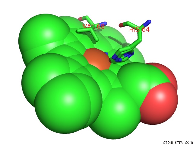
Mono view
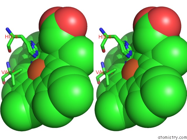
Stereo pair view
Mono view
Stereo pair view
A full contact list of Iron with other atoms in the Fe binding
site number 1 of Crystal Structure of the Oxy Myoglobin Free From X-Ray- Induced Photoreduction within 5.0Å range:
|
Reference:
M.Unno,
H.Chen,
S.Kusama,
S.Shaik,
M.Ikeda-Saito.
Structural Characterization of the Fleeting Ferric Peroxo Species in Myoglobin: Experiment and Theory J.Am.Chem.Soc. V. 129 13394 2007.
ISSN: ISSN 0002-7863
PubMed: 17929929
DOI: 10.1021/JA076108X
Page generated: Sun Aug 4 05:54:52 2024
ISSN: ISSN 0002-7863
PubMed: 17929929
DOI: 10.1021/JA076108X
Last articles
Zn in 9J0NZn in 9J0O
Zn in 9J0P
Zn in 9FJX
Zn in 9EKB
Zn in 9C0F
Zn in 9CAH
Zn in 9CH0
Zn in 9CH3
Zn in 9CH1