Iron »
PDB 2z5z-2zpg »
2zax »
Iron in PDB 2zax: Crystal Structure of Ferric Cytochrome P450CAM
Enzymatic activity of Crystal Structure of Ferric Cytochrome P450CAM
All present enzymatic activity of Crystal Structure of Ferric Cytochrome P450CAM:
1.14.15.1;
1.14.15.1;
Protein crystallography data
The structure of Crystal Structure of Ferric Cytochrome P450CAM, PDB code: 2zax
was solved by
K.Sakurai,
H.Shimada,
K.Harada,
T.Hayashi,
T.Tsukihara,
with X-Ray Crystallography technique. A brief refinement statistics is given in the table below:
| Resolution Low / High (Å) | 31.77 / 1.60 |
| Space group | P 43 21 2 |
| Cell size a, b, c (Å), α, β, γ (°) | 63.522, 63.522, 249.670, 90.00, 90.00, 90.00 |
| R / Rfree (%) | 16.5 / 18.3 |
Other elements in 2zax:
The structure of Crystal Structure of Ferric Cytochrome P450CAM also contains other interesting chemical elements:
| Potassium | (K) | 1 atom |
Iron Binding Sites:
The binding sites of Iron atom in the Crystal Structure of Ferric Cytochrome P450CAM
(pdb code 2zax). This binding sites where shown within
5.0 Angstroms radius around Iron atom.
In total only one binding site of Iron was determined in the Crystal Structure of Ferric Cytochrome P450CAM, PDB code: 2zax:
In total only one binding site of Iron was determined in the Crystal Structure of Ferric Cytochrome P450CAM, PDB code: 2zax:
Iron binding site 1 out of 1 in 2zax
Go back to
Iron binding site 1 out
of 1 in the Crystal Structure of Ferric Cytochrome P450CAM
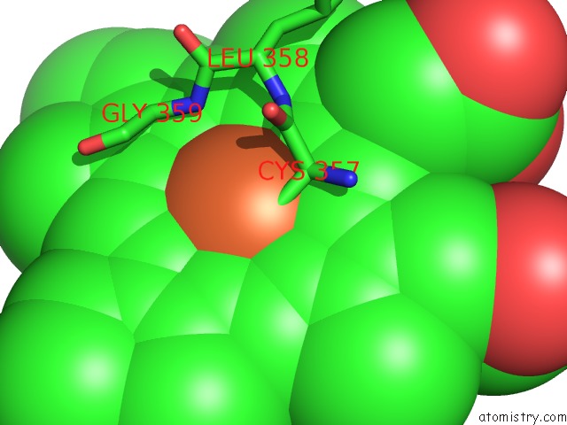
Mono view
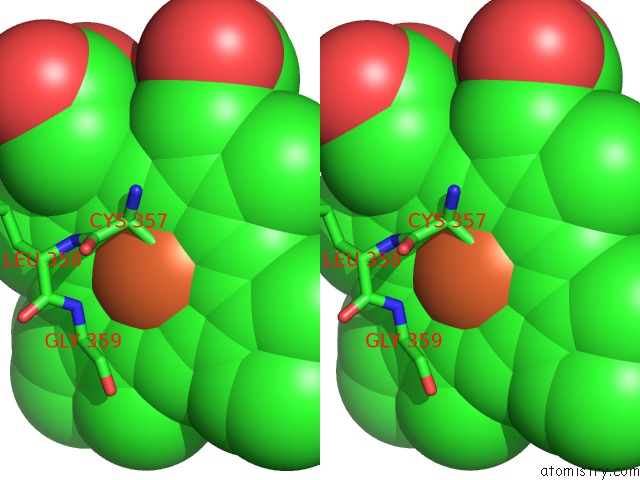
Stereo pair view
Mono view
Stereo pair view
A full contact list of Iron with other atoms in the Fe binding
site number 1 of Crystal Structure of Ferric Cytochrome P450CAM within 5.0Å range:
|
Reference:
K.Harada,
K.Sakurai,
K.Ikemura,
T.Ogura,
S.Hirota,
H.Shimada,
T.Hayashi.
Evaluation of the Functional Role of the Heme-6-Propionate Side Chain in Cytochrome P450CAM J.Am.Chem.Soc. V. 130 432 2008.
ISSN: ISSN 0002-7863
PubMed: 18088124
DOI: 10.1021/JA077902L
Page generated: Sun Aug 4 05:56:17 2024
ISSN: ISSN 0002-7863
PubMed: 18088124
DOI: 10.1021/JA077902L
Last articles
Fe in 2YXOFe in 2YRS
Fe in 2YXC
Fe in 2YNM
Fe in 2YVJ
Fe in 2YP1
Fe in 2YU2
Fe in 2YU1
Fe in 2YQB
Fe in 2YOO