Iron »
PDB 2z5z-2zpg »
2zlt »
Iron in PDB 2zlt: Horse Methemoglobin High Salt, pH 7.0
Protein crystallography data
The structure of Horse Methemoglobin High Salt, pH 7.0, PDB code: 2zlt
was solved by
P.S.Kaushal,
R.Sankaranarayanan,
M.Vijayan,
with X-Ray Crystallography technique. A brief refinement statistics is given in the table below:
| Resolution Low / High (Å) | 18.32 / 1.90 |
| Space group | C 1 2 1 |
| Cell size a, b, c (Å), α, β, γ (°) | 108.650, 63.336, 54.957, 90.00, 110.84, 90.00 |
| R / Rfree (%) | 19 / 22.6 |
Iron Binding Sites:
The binding sites of Iron atom in the Horse Methemoglobin High Salt, pH 7.0
(pdb code 2zlt). This binding sites where shown within
5.0 Angstroms radius around Iron atom.
In total 2 binding sites of Iron where determined in the Horse Methemoglobin High Salt, pH 7.0, PDB code: 2zlt:
Jump to Iron binding site number: 1; 2;
In total 2 binding sites of Iron where determined in the Horse Methemoglobin High Salt, pH 7.0, PDB code: 2zlt:
Jump to Iron binding site number: 1; 2;
Iron binding site 1 out of 2 in 2zlt
Go back to
Iron binding site 1 out
of 2 in the Horse Methemoglobin High Salt, pH 7.0
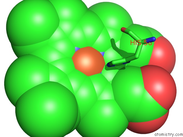
Mono view
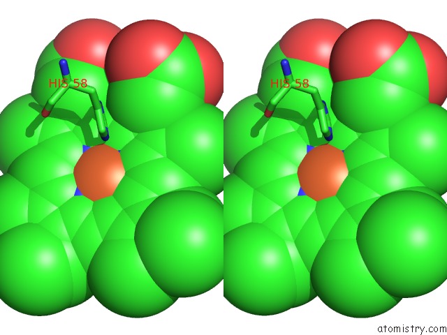
Stereo pair view
Mono view
Stereo pair view
A full contact list of Iron with other atoms in the Fe binding
site number 1 of Horse Methemoglobin High Salt, pH 7.0 within 5.0Å range:
|
Iron binding site 2 out of 2 in 2zlt
Go back to
Iron binding site 2 out
of 2 in the Horse Methemoglobin High Salt, pH 7.0
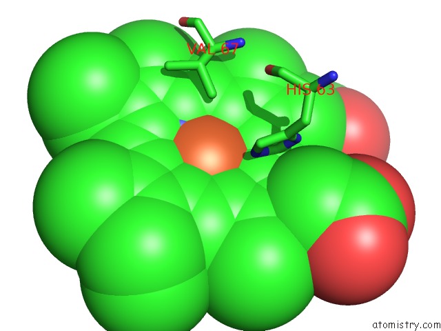
Mono view
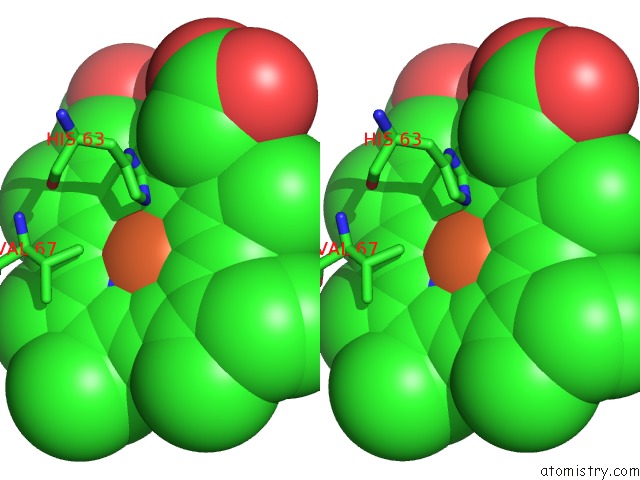
Stereo pair view
Mono view
Stereo pair view
A full contact list of Iron with other atoms in the Fe binding
site number 2 of Horse Methemoglobin High Salt, pH 7.0 within 5.0Å range:
|
Reference:
P.S.Kaushal,
R.Sankaranarayanan,
M.Vijayan.
Water-Mediated Variability in the Structure of Relaxed-State Haemoglobin Acta Crystallogr.,Sect.F V. 64 463 2008.
ISSN: ESSN 1744-3091
PubMed: 18540052
DOI: 10.1107/S1744309108013109
Page generated: Sun Aug 4 05:59:50 2024
ISSN: ESSN 1744-3091
PubMed: 18540052
DOI: 10.1107/S1744309108013109
Last articles
Zn in 9J0NZn in 9J0O
Zn in 9J0P
Zn in 9FJX
Zn in 9EKB
Zn in 9C0F
Zn in 9CAH
Zn in 9CH0
Zn in 9CH3
Zn in 9CH1