Iron »
PDB 2zph-3a0b »
2zvs »
Iron in PDB 2zvs: Crystal Structure of the 2[4FE-4S] Ferredoxin From Escherichia Coli
Protein crystallography data
The structure of Crystal Structure of the 2[4FE-4S] Ferredoxin From Escherichia Coli, PDB code: 2zvs
was solved by
P.Giastas,
M.I.Mavridis,
with X-Ray Crystallography technique. A brief refinement statistics is given in the table below:
| Resolution Low / High (Å) | 30.00 / 1.65 |
| Space group | P 62 |
| Cell size a, b, c (Å), α, β, γ (°) | 65.543, 65.543, 132.367, 90.00, 90.00, 120.00 |
| R / Rfree (%) | 21.1 / 27.3 |
Iron Binding Sites:
Pages:
>>> Page 1 <<< Page 2, Binding sites: 11 - 20; Page 3, Binding sites: 21 - 24;Binding sites:
The binding sites of Iron atom in the Crystal Structure of the 2[4FE-4S] Ferredoxin From Escherichia Coli (pdb code 2zvs). This binding sites where shown within 5.0 Angstroms radius around Iron atom.In total 24 binding sites of Iron where determined in the Crystal Structure of the 2[4FE-4S] Ferredoxin From Escherichia Coli, PDB code: 2zvs:
Jump to Iron binding site number: 1; 2; 3; 4; 5; 6; 7; 8; 9; 10;
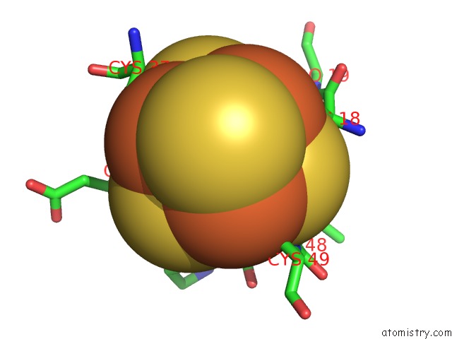
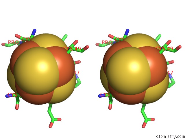
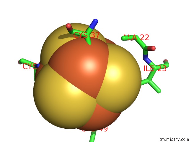
Iron binding site 1 out of 24 in 2zvs
Go back to
Iron binding site 1 out
of 24 in the Crystal Structure of the 2[4FE-4S] Ferredoxin From Escherichia Coli

Mono view
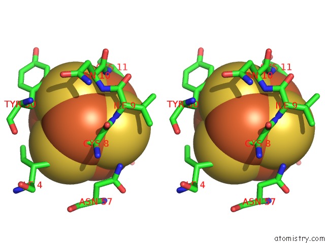
Stereo pair view
Mono view
Stereo pair view
A full contact list of Iron with other atoms in the Fe binding
site number 1 of Crystal Structure of the 2[4FE-4S] Ferredoxin From Escherichia Coli within 5.0Å range:
|
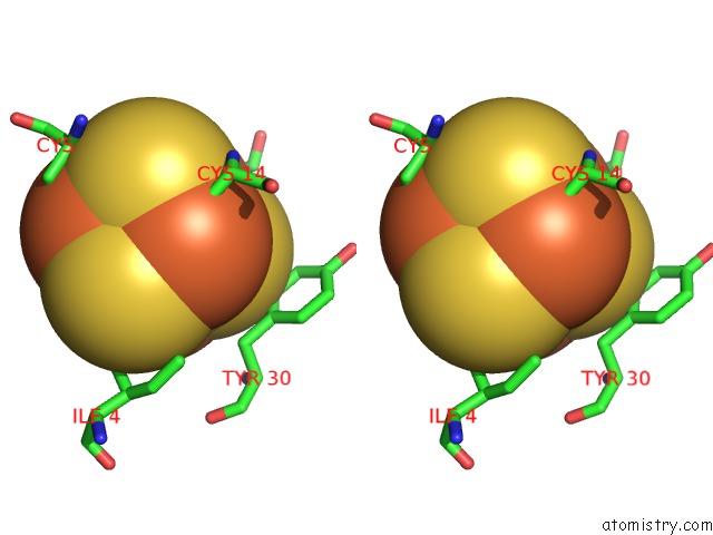
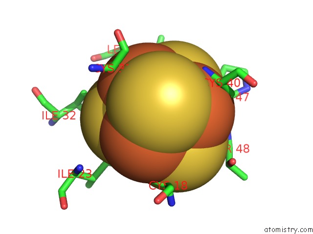
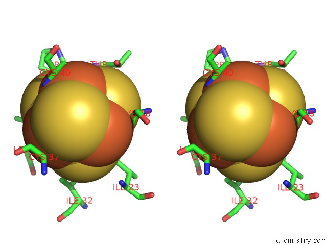
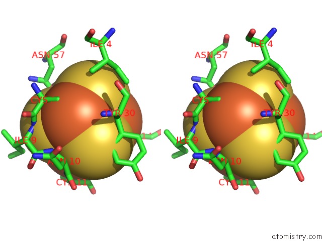
Iron binding site 2 out of 24 in 2zvs
Go back to
Iron binding site 2 out
of 24 in the Crystal Structure of the 2[4FE-4S] Ferredoxin From Escherichia Coli

Mono view

Stereo pair view
Mono view
Stereo pair view
A full contact list of Iron with other atoms in the Fe binding
site number 2 of Crystal Structure of the 2[4FE-4S] Ferredoxin From Escherichia Coli within 5.0Å range:
|
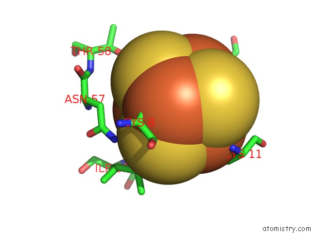
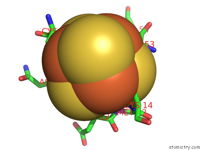
Iron binding site 3 out of 24 in 2zvs
Go back to
Iron binding site 3 out
of 24 in the Crystal Structure of the 2[4FE-4S] Ferredoxin From Escherichia Coli

Mono view
Stereo pair view
Mono view
Stereo pair view
A full contact list of Iron with other atoms in the Fe binding
site number 3 of Crystal Structure of the 2[4FE-4S] Ferredoxin From Escherichia Coli within 5.0Å range:
|
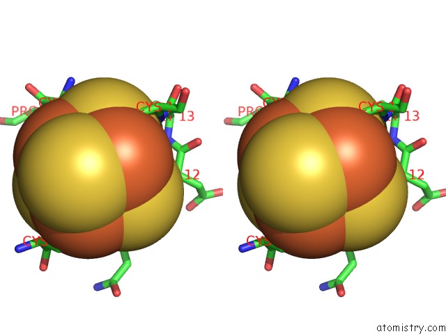
Iron binding site 4 out of 24 in 2zvs
Go back to
Iron binding site 4 out
of 24 in the Crystal Structure of the 2[4FE-4S] Ferredoxin From Escherichia Coli
Mono view
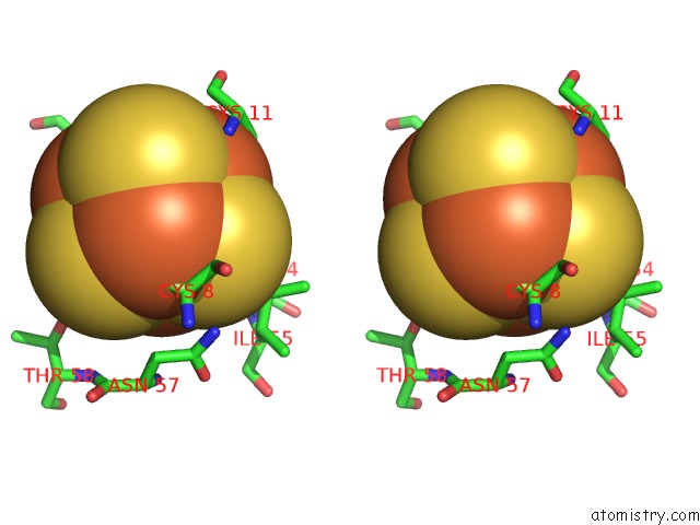
Stereo pair view
Mono view
Stereo pair view
A full contact list of Iron with other atoms in the Fe binding
site number 4 of Crystal Structure of the 2[4FE-4S] Ferredoxin From Escherichia Coli within 5.0Å range:
|
Iron binding site 5 out of 24 in 2zvs
Go back to
Iron binding site 5 out
of 24 in the Crystal Structure of the 2[4FE-4S] Ferredoxin From Escherichia Coli

Mono view

Stereo pair view
Mono view
Stereo pair view
A full contact list of Iron with other atoms in the Fe binding
site number 5 of Crystal Structure of the 2[4FE-4S] Ferredoxin From Escherichia Coli within 5.0Å range:
|
Iron binding site 6 out of 24 in 2zvs
Go back to
Iron binding site 6 out
of 24 in the Crystal Structure of the 2[4FE-4S] Ferredoxin From Escherichia Coli
Mono view
Stereo pair view
Mono view
Stereo pair view
A full contact list of Iron with other atoms in the Fe binding
site number 6 of Crystal Structure of the 2[4FE-4S] Ferredoxin From Escherichia Coli within 5.0Å range:
|
Iron binding site 7 out of 24 in 2zvs
Go back to
Iron binding site 7 out
of 24 in the Crystal Structure of the 2[4FE-4S] Ferredoxin From Escherichia Coli
Mono view
Stereo pair view
Mono view
Stereo pair view
A full contact list of Iron with other atoms in the Fe binding
site number 7 of Crystal Structure of the 2[4FE-4S] Ferredoxin From Escherichia Coli within 5.0Å range:
|
Iron binding site 8 out of 24 in 2zvs
Go back to
Iron binding site 8 out
of 24 in the Crystal Structure of the 2[4FE-4S] Ferredoxin From Escherichia Coli
Mono view

Stereo pair view
Mono view
Stereo pair view
A full contact list of Iron with other atoms in the Fe binding
site number 8 of Crystal Structure of the 2[4FE-4S] Ferredoxin From Escherichia Coli within 5.0Å range:
|
Iron binding site 9 out of 24 in 2zvs
Go back to
Iron binding site 9 out
of 24 in the Crystal Structure of the 2[4FE-4S] Ferredoxin From Escherichia Coli

Mono view
Stereo pair view
Mono view
Stereo pair view
A full contact list of Iron with other atoms in the Fe binding
site number 9 of Crystal Structure of the 2[4FE-4S] Ferredoxin From Escherichia Coli within 5.0Å range:
|
Iron binding site 10 out of 24 in 2zvs
Go back to
Iron binding site 10 out
of 24 in the Crystal Structure of the 2[4FE-4S] Ferredoxin From Escherichia Coli
Mono view
Stereo pair view
Mono view
Stereo pair view
A full contact list of Iron with other atoms in the Fe binding
site number 10 of Crystal Structure of the 2[4FE-4S] Ferredoxin From Escherichia Coli within 5.0Å range:
|
Reference:
E.Saridakis,
P.Giastas,
G.Efthymiou,
V.Thoma,
J.M.Moulis,
P.Kyritsis,
I.M.Mavridis.
Insight Into the Protein and Solvent Contributions to the Reduction Potentials of [4FE-4S]2+/+ Clusters: Crystal Structures of the Allochromatium Vinosum Ferredoxin Variants C57A and V13G and the Homologous Escherichia Coli Ferredoxin J.Biol.Inorg.Chem. V. 14 783 2009.
ISSN: ISSN 0949-8257
PubMed: 19290553
DOI: 10.1007/S00775-009-0492-X
Page generated: Sun Aug 4 06:23:21 2024
ISSN: ISSN 0949-8257
PubMed: 19290553
DOI: 10.1007/S00775-009-0492-X
Last articles
Zn in 9MJ5Zn in 9HNW
Zn in 9G0L
Zn in 9FNE
Zn in 9DZN
Zn in 9E0I
Zn in 9D32
Zn in 9DAK
Zn in 8ZXC
Zn in 8ZUF