Iron »
PDB 2zph-3a0b »
2zyl »
Iron in PDB 2zyl: Crystal Structure of 3-Ketosteroid-9-Alpha-Hydroxylase (Ksha) From M. Tuberculosis
Protein crystallography data
The structure of Crystal Structure of 3-Ketosteroid-9-Alpha-Hydroxylase (Ksha) From M. Tuberculosis, PDB code: 2zyl
was solved by
I.D'angelo,
J.Capyk,
N.Strynadka,
L.Eltis,
with X-Ray Crystallography technique. A brief refinement statistics is given in the table below:
| Resolution Low / High (Å) | 19.85 / 2.30 |
| Space group | P 3 2 1 |
| Cell size a, b, c (Å), α, β, γ (°) | 116.200, 116.200, 81.000, 90.00, 90.00, 120.00 |
| R / Rfree (%) | 19.7 / 23.5 |
Other elements in 2zyl:
The structure of Crystal Structure of 3-Ketosteroid-9-Alpha-Hydroxylase (Ksha) From M. Tuberculosis also contains other interesting chemical elements:
| Sodium | (Na) | 1 atom |
Iron Binding Sites:
The binding sites of Iron atom in the Crystal Structure of 3-Ketosteroid-9-Alpha-Hydroxylase (Ksha) From M. Tuberculosis
(pdb code 2zyl). This binding sites where shown within
5.0 Angstroms radius around Iron atom.
In total 3 binding sites of Iron where determined in the Crystal Structure of 3-Ketosteroid-9-Alpha-Hydroxylase (Ksha) From M. Tuberculosis, PDB code: 2zyl:
Jump to Iron binding site number: 1; 2; 3;
In total 3 binding sites of Iron where determined in the Crystal Structure of 3-Ketosteroid-9-Alpha-Hydroxylase (Ksha) From M. Tuberculosis, PDB code: 2zyl:
Jump to Iron binding site number: 1; 2; 3;
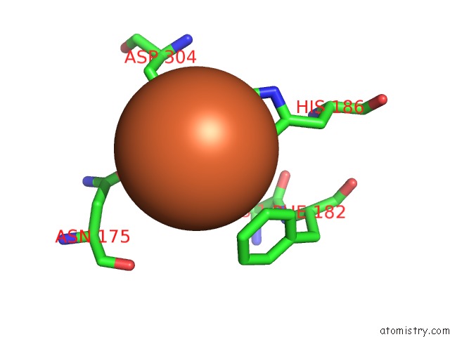
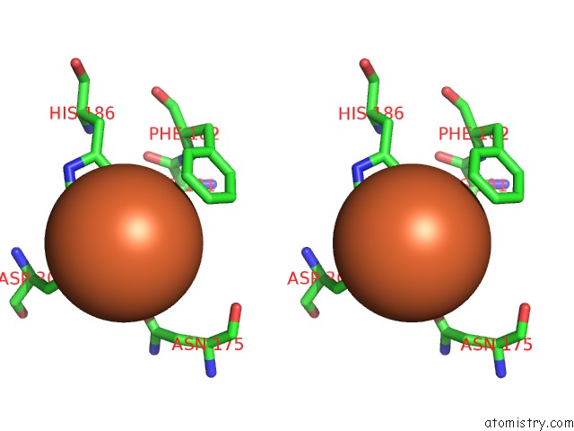
Iron binding site 1 out of 3 in 2zyl
Go back to
Iron binding site 1 out
of 3 in the Crystal Structure of 3-Ketosteroid-9-Alpha-Hydroxylase (Ksha) From M. Tuberculosis
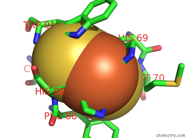
Mono view
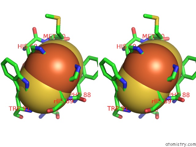
Stereo pair view
Mono view
Stereo pair view
A full contact list of Iron with other atoms in the Fe binding
site number 1 of Crystal Structure of 3-Ketosteroid-9-Alpha-Hydroxylase (Ksha) From M. Tuberculosis within 5.0Å range:
|
Iron binding site 2 out of 3 in 2zyl
Go back to
Iron binding site 2 out
of 3 in the Crystal Structure of 3-Ketosteroid-9-Alpha-Hydroxylase (Ksha) From M. Tuberculosis
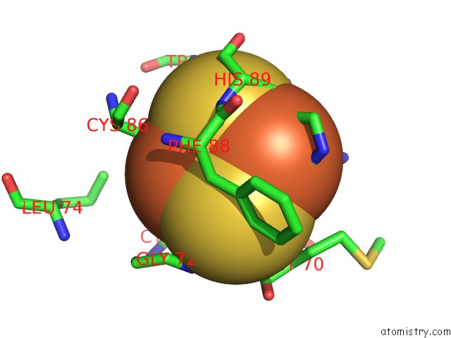
Mono view
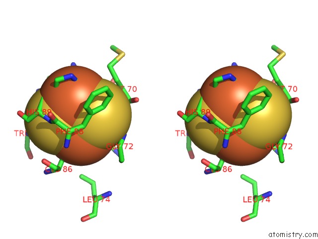
Stereo pair view
Mono view
Stereo pair view
A full contact list of Iron with other atoms in the Fe binding
site number 2 of Crystal Structure of 3-Ketosteroid-9-Alpha-Hydroxylase (Ksha) From M. Tuberculosis within 5.0Å range:
|
Iron binding site 3 out of 3 in 2zyl
Go back to
Iron binding site 3 out
of 3 in the Crystal Structure of 3-Ketosteroid-9-Alpha-Hydroxylase (Ksha) From M. Tuberculosis
Mono view
Stereo pair view
Mono view
Stereo pair view
A full contact list of Iron with other atoms in the Fe binding
site number 3 of Crystal Structure of 3-Ketosteroid-9-Alpha-Hydroxylase (Ksha) From M. Tuberculosis within 5.0Å range:
|
Reference:
J.K.Capyk,
I.D'angelo,
N.C.Strynadka,
L.D.Eltis.
Characterization of 3-Ketosteroid 9{Alpha}-Hydroxylase, A Rieske Oxygenase in the Cholesterol Degradation Pathway of Mycobacterium Tuberculosis J.Biol.Chem. V. 284 9937 2009.
ISSN: ISSN 0021-9258
PubMed: 19234303
DOI: 10.1074/JBC.M900719200
Page generated: Sun Aug 4 06:24:52 2024
ISSN: ISSN 0021-9258
PubMed: 19234303
DOI: 10.1074/JBC.M900719200
Last articles
Zn in 9J0NZn in 9J0O
Zn in 9J0P
Zn in 9FJX
Zn in 9EKB
Zn in 9C0F
Zn in 9CAH
Zn in 9CH0
Zn in 9CH3
Zn in 9CH1