Iron »
PDB 3a0g-3ae5 »
3a8h »
Iron in PDB 3a8h: Crystal Structure of Nitrile Hydratase Mutant S113A Complexed with Trimethylacetamide
Enzymatic activity of Crystal Structure of Nitrile Hydratase Mutant S113A Complexed with Trimethylacetamide
All present enzymatic activity of Crystal Structure of Nitrile Hydratase Mutant S113A Complexed with Trimethylacetamide:
4.2.1.84;
4.2.1.84;
Protein crystallography data
The structure of Crystal Structure of Nitrile Hydratase Mutant S113A Complexed with Trimethylacetamide, PDB code: 3a8h
was solved by
Y.Yamanaka,
K.Hashimoto,
A.Ohtaki,
K.Noguchi,
M.Yohda,
M.Odaka,
with X-Ray Crystallography technique. A brief refinement statistics is given in the table below:
| Resolution Low / High (Å) | 28.09 / 1.66 |
| Space group | C 1 2 1 |
| Cell size a, b, c (Å), α, β, γ (°) | 113.606, 60.012, 81.499, 90.00, 125.14, 90.00 |
| R / Rfree (%) | 17.3 / 20 |
Iron Binding Sites:
The binding sites of Iron atom in the Crystal Structure of Nitrile Hydratase Mutant S113A Complexed with Trimethylacetamide
(pdb code 3a8h). This binding sites where shown within
5.0 Angstroms radius around Iron atom.
In total only one binding site of Iron was determined in the Crystal Structure of Nitrile Hydratase Mutant S113A Complexed with Trimethylacetamide, PDB code: 3a8h:
In total only one binding site of Iron was determined in the Crystal Structure of Nitrile Hydratase Mutant S113A Complexed with Trimethylacetamide, PDB code: 3a8h:
Iron binding site 1 out of 1 in 3a8h
Go back to
Iron binding site 1 out
of 1 in the Crystal Structure of Nitrile Hydratase Mutant S113A Complexed with Trimethylacetamide
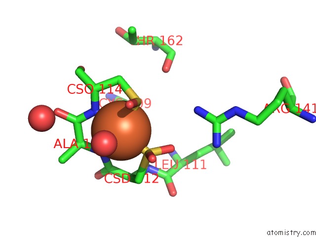
Mono view
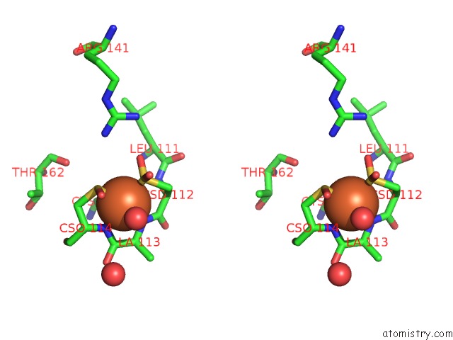
Stereo pair view
Mono view
Stereo pair view
A full contact list of Iron with other atoms in the Fe binding
site number 1 of Crystal Structure of Nitrile Hydratase Mutant S113A Complexed with Trimethylacetamide within 5.0Å range:
|
Reference:
Y.Yamanaka,
K.Hashimoto,
A.Ohtaki,
K.Noguchi,
M.Yohda,
M.Odaka.
Kinetic and Structural Studies on Roles of the Serine Ligand and A Strictly Conserved Tyrosine Residue in Nitrile Hydratase J.Biol.Inorg.Chem. V. 15 655 2010.
ISSN: ISSN 0949-8257
PubMed: 20221653
DOI: 10.1007/S00775-010-0632-3
Page generated: Sun Aug 4 06:44:30 2024
ISSN: ISSN 0949-8257
PubMed: 20221653
DOI: 10.1007/S00775-010-0632-3
Last articles
Zn in 9MJ5Zn in 9HNW
Zn in 9G0L
Zn in 9FNE
Zn in 9DZN
Zn in 9E0I
Zn in 9D32
Zn in 9DAK
Zn in 8ZXC
Zn in 8ZUF