Iron »
PDB 3a0g-3ae5 »
3a9m »
Iron in PDB 3a9m: Crystal Structure of A Hemoglobin Component V From Propsilocerus Akamusi (PH9.0 Coordinates)
Protein crystallography data
The structure of Crystal Structure of A Hemoglobin Component V From Propsilocerus Akamusi (PH9.0 Coordinates), PDB code: 3a9m
was solved by
T.Kuwada,
T.Hasegawa,
T.Takagi,
F.Shishikura,
with X-Ray Crystallography technique. A brief refinement statistics is given in the table below:
| Resolution Low / High (Å) | 24.14 / 1.80 |
| Space group | P 21 21 21 |
| Cell size a, b, c (Å), α, β, γ (°) | 33.580, 64.510, 72.820, 90.00, 90.00, 90.00 |
| R / Rfree (%) | 21.4 / 24.3 |
Iron Binding Sites:
The binding sites of Iron atom in the Crystal Structure of A Hemoglobin Component V From Propsilocerus Akamusi (PH9.0 Coordinates)
(pdb code 3a9m). This binding sites where shown within
5.0 Angstroms radius around Iron atom.
In total only one binding site of Iron was determined in the Crystal Structure of A Hemoglobin Component V From Propsilocerus Akamusi (PH9.0 Coordinates), PDB code: 3a9m:
In total only one binding site of Iron was determined in the Crystal Structure of A Hemoglobin Component V From Propsilocerus Akamusi (PH9.0 Coordinates), PDB code: 3a9m:
Iron binding site 1 out of 1 in 3a9m
Go back to
Iron binding site 1 out
of 1 in the Crystal Structure of A Hemoglobin Component V From Propsilocerus Akamusi (PH9.0 Coordinates)
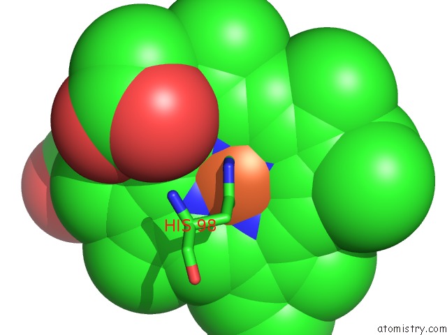
Mono view
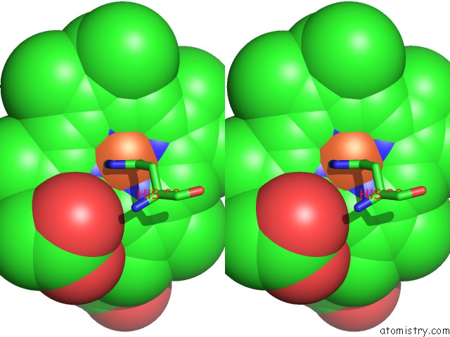
Stereo pair view
Mono view
Stereo pair view
A full contact list of Iron with other atoms in the Fe binding
site number 1 of Crystal Structure of A Hemoglobin Component V From Propsilocerus Akamusi (PH9.0 Coordinates) within 5.0Å range:
|
Reference:
T.Kuwada,
T.Hasegawa,
T.Takagi,
I.Sato,
F.Shishikura.
pH-Dependent Structural Changes in Haemoglobin Component V From the Midge Larva Propsilocerus Akamusi (Orthocladiinae, Diptera) Acta Crystallogr.,Sect.D V. 66 258 2010.
ISSN: ISSN 0907-4449
PubMed: 20179337
DOI: 10.1107/S0907444909055760
Page generated: Sun Aug 4 06:45:13 2024
ISSN: ISSN 0907-4449
PubMed: 20179337
DOI: 10.1107/S0907444909055760
Last articles
Zn in 9MJ5Zn in 9HNW
Zn in 9G0L
Zn in 9FNE
Zn in 9DZN
Zn in 9E0I
Zn in 9D32
Zn in 9DAK
Zn in 8ZXC
Zn in 8ZUF