Iron »
PDB 3ae6-3arj »
3aer »
Iron in PDB 3aer: Structure of the Light-Independent Protochlorophyllide Reductase Catalyzing A Key Reduction For Greening in the Dark
Protein crystallography data
The structure of Structure of the Light-Independent Protochlorophyllide Reductase Catalyzing A Key Reduction For Greening in the Dark, PDB code: 3aer
was solved by
N.Muraki,
J.Nomata,
T.Shiba,
Y.Fujita,
G.Kurisu,
with X-Ray Crystallography technique. A brief refinement statistics is given in the table below:
| Resolution Low / High (Å) | 47.09 / 2.80 |
| Space group | P 1 21 1 |
| Cell size a, b, c (Å), α, β, γ (°) | 81.418, 80.941, 176.728, 90.00, 100.66, 90.00 |
| R / Rfree (%) | 18.7 / 23.7 |
Iron Binding Sites:
The binding sites of Iron atom in the Structure of the Light-Independent Protochlorophyllide Reductase Catalyzing A Key Reduction For Greening in the Dark
(pdb code 3aer). This binding sites where shown within
5.0 Angstroms radius around Iron atom.
In total 8 binding sites of Iron where determined in the Structure of the Light-Independent Protochlorophyllide Reductase Catalyzing A Key Reduction For Greening in the Dark, PDB code: 3aer:
Jump to Iron binding site number: 1; 2; 3; 4; 5; 6; 7; 8;
In total 8 binding sites of Iron where determined in the Structure of the Light-Independent Protochlorophyllide Reductase Catalyzing A Key Reduction For Greening in the Dark, PDB code: 3aer:
Jump to Iron binding site number: 1; 2; 3; 4; 5; 6; 7; 8;
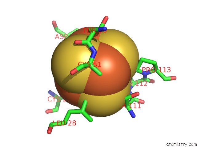
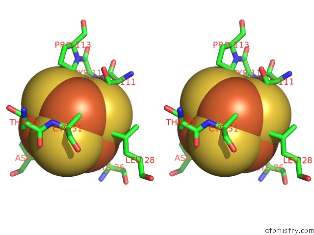
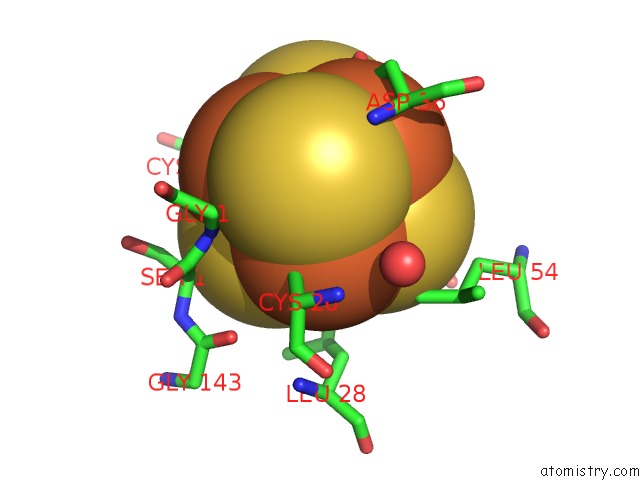
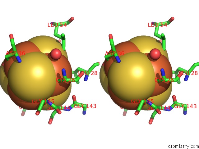
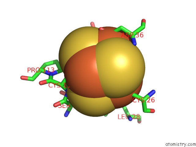
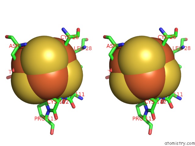
Iron binding site 1 out of 8 in 3aer
Go back to
Iron binding site 1 out
of 8 in the Structure of the Light-Independent Protochlorophyllide Reductase Catalyzing A Key Reduction For Greening in the Dark
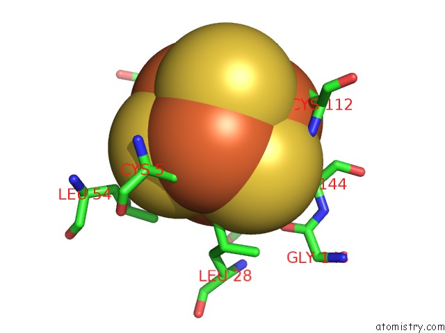
Mono view
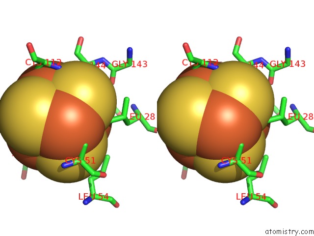
Stereo pair view
Mono view
Stereo pair view
A full contact list of Iron with other atoms in the Fe binding
site number 1 of Structure of the Light-Independent Protochlorophyllide Reductase Catalyzing A Key Reduction For Greening in the Dark within 5.0Å range:
|
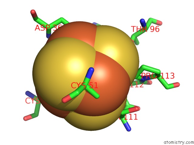
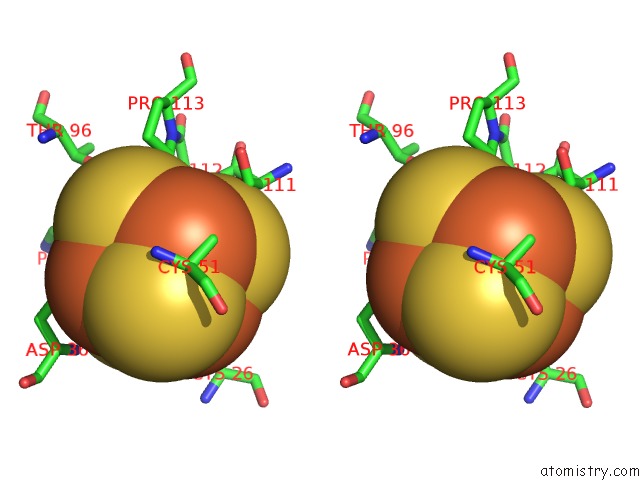
Iron binding site 2 out of 8 in 3aer
Go back to
Iron binding site 2 out
of 8 in the Structure of the Light-Independent Protochlorophyllide Reductase Catalyzing A Key Reduction For Greening in the Dark
Mono view
Stereo pair view
Mono view
Stereo pair view
A full contact list of Iron with other atoms in the Fe binding
site number 2 of Structure of the Light-Independent Protochlorophyllide Reductase Catalyzing A Key Reduction For Greening in the Dark within 5.0Å range:
|
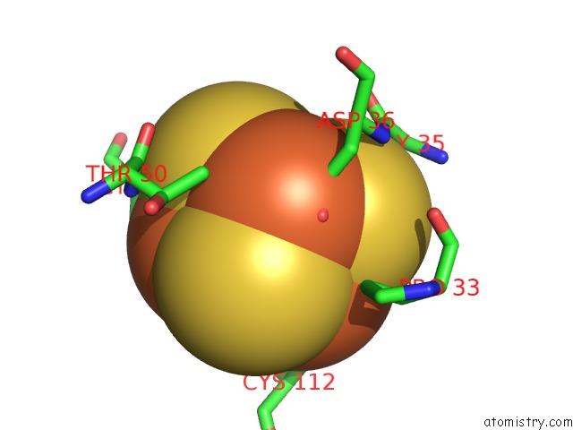
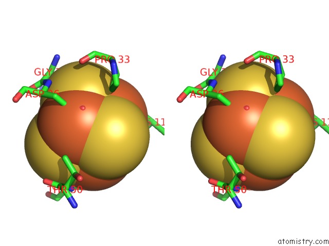
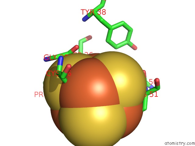
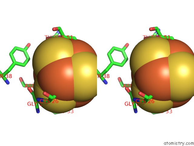
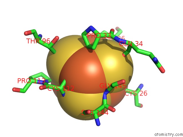
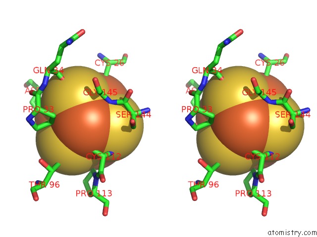
Iron binding site 3 out of 8 in 3aer
Go back to
Iron binding site 3 out
of 8 in the Structure of the Light-Independent Protochlorophyllide Reductase Catalyzing A Key Reduction For Greening in the Dark
Mono view
Stereo pair view
Mono view
Stereo pair view
A full contact list of Iron with other atoms in the Fe binding
site number 3 of Structure of the Light-Independent Protochlorophyllide Reductase Catalyzing A Key Reduction For Greening in the Dark within 5.0Å range:
|
Iron binding site 4 out of 8 in 3aer
Go back to
Iron binding site 4 out
of 8 in the Structure of the Light-Independent Protochlorophyllide Reductase Catalyzing A Key Reduction For Greening in the Dark
Mono view
Stereo pair view
Mono view
Stereo pair view
A full contact list of Iron with other atoms in the Fe binding
site number 4 of Structure of the Light-Independent Protochlorophyllide Reductase Catalyzing A Key Reduction For Greening in the Dark within 5.0Å range:
|
Iron binding site 5 out of 8 in 3aer
Go back to
Iron binding site 5 out
of 8 in the Structure of the Light-Independent Protochlorophyllide Reductase Catalyzing A Key Reduction For Greening in the Dark
Mono view
Stereo pair view
Mono view
Stereo pair view
A full contact list of Iron with other atoms in the Fe binding
site number 5 of Structure of the Light-Independent Protochlorophyllide Reductase Catalyzing A Key Reduction For Greening in the Dark within 5.0Å range:
|
Iron binding site 6 out of 8 in 3aer
Go back to
Iron binding site 6 out
of 8 in the Structure of the Light-Independent Protochlorophyllide Reductase Catalyzing A Key Reduction For Greening in the Dark
Mono view
Stereo pair view
Mono view
Stereo pair view
A full contact list of Iron with other atoms in the Fe binding
site number 6 of Structure of the Light-Independent Protochlorophyllide Reductase Catalyzing A Key Reduction For Greening in the Dark within 5.0Å range:
|
Iron binding site 7 out of 8 in 3aer
Go back to
Iron binding site 7 out
of 8 in the Structure of the Light-Independent Protochlorophyllide Reductase Catalyzing A Key Reduction For Greening in the Dark
Mono view
Stereo pair view
Mono view
Stereo pair view
A full contact list of Iron with other atoms in the Fe binding
site number 7 of Structure of the Light-Independent Protochlorophyllide Reductase Catalyzing A Key Reduction For Greening in the Dark within 5.0Å range:
|
Iron binding site 8 out of 8 in 3aer
Go back to
Iron binding site 8 out
of 8 in the Structure of the Light-Independent Protochlorophyllide Reductase Catalyzing A Key Reduction For Greening in the Dark
Mono view
Stereo pair view
Mono view
Stereo pair view
A full contact list of Iron with other atoms in the Fe binding
site number 8 of Structure of the Light-Independent Protochlorophyllide Reductase Catalyzing A Key Reduction For Greening in the Dark within 5.0Å range:
|
Reference:
N.Muraki,
J.Nomata,
K.Ebata,
T.Mizoguchi,
T.Shiba,
H.Tamiaki,
G.Kurisu,
Y.Fujita.
X-Ray Crystal Structure of the Light-Independent Protochlorophyllide Reductase Nature V. 465 110 2010.
ISSN: ISSN 0028-0836
PubMed: 20400946
DOI: 10.1038/NATURE08950
Page generated: Sun Aug 4 07:07:39 2024
ISSN: ISSN 0028-0836
PubMed: 20400946
DOI: 10.1038/NATURE08950
Last articles
Zn in 9J0NZn in 9J0O
Zn in 9J0P
Zn in 9FJX
Zn in 9EKB
Zn in 9C0F
Zn in 9CAH
Zn in 9CH0
Zn in 9CH3
Zn in 9CH1