Iron »
PDB 3ae6-3arj »
3aqj »
Iron in PDB 3aqj: Crystal Structure of A C-Terminal Domain of the Bacteriophage P2 Tail Spike Protein, Gpv
Protein crystallography data
The structure of Crystal Structure of A C-Terminal Domain of the Bacteriophage P2 Tail Spike Protein, Gpv, PDB code: 3aqj
was solved by
S.Takeda,
E.Yamashita,
A.Nakagawa,
with X-Ray Crystallography technique. A brief refinement statistics is given in the table below:
| Resolution Low / High (Å) | 46.42 / 1.27 |
| Space group | P 1 21 1 |
| Cell size a, b, c (Å), α, β, γ (°) | 66.912, 64.463, 67.186, 90.00, 91.32, 90.00 |
| R / Rfree (%) | 16 / 19 |
Other elements in 3aqj:
The structure of Crystal Structure of A C-Terminal Domain of the Bacteriophage P2 Tail Spike Protein, Gpv also contains other interesting chemical elements:
| Calcium | (Ca) | 2 atoms |
| Chlorine | (Cl) | 2 atoms |
Iron Binding Sites:
The binding sites of Iron atom in the Crystal Structure of A C-Terminal Domain of the Bacteriophage P2 Tail Spike Protein, Gpv
(pdb code 3aqj). This binding sites where shown within
5.0 Angstroms radius around Iron atom.
In total 2 binding sites of Iron where determined in the Crystal Structure of A C-Terminal Domain of the Bacteriophage P2 Tail Spike Protein, Gpv, PDB code: 3aqj:
Jump to Iron binding site number: 1; 2;
In total 2 binding sites of Iron where determined in the Crystal Structure of A C-Terminal Domain of the Bacteriophage P2 Tail Spike Protein, Gpv, PDB code: 3aqj:
Jump to Iron binding site number: 1; 2;
Iron binding site 1 out of 2 in 3aqj
Go back to
Iron binding site 1 out
of 2 in the Crystal Structure of A C-Terminal Domain of the Bacteriophage P2 Tail Spike Protein, Gpv
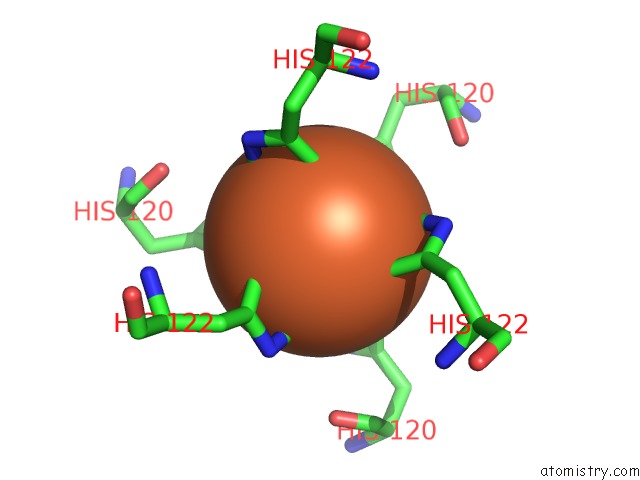
Mono view
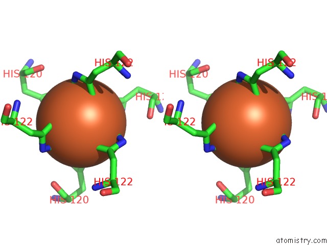
Stereo pair view
Mono view
Stereo pair view
A full contact list of Iron with other atoms in the Fe binding
site number 1 of Crystal Structure of A C-Terminal Domain of the Bacteriophage P2 Tail Spike Protein, Gpv within 5.0Å range:
|
Iron binding site 2 out of 2 in 3aqj
Go back to
Iron binding site 2 out
of 2 in the Crystal Structure of A C-Terminal Domain of the Bacteriophage P2 Tail Spike Protein, Gpv
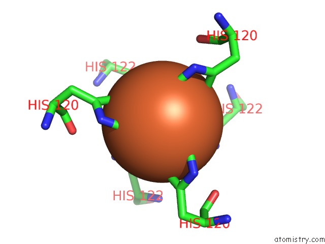
Mono view
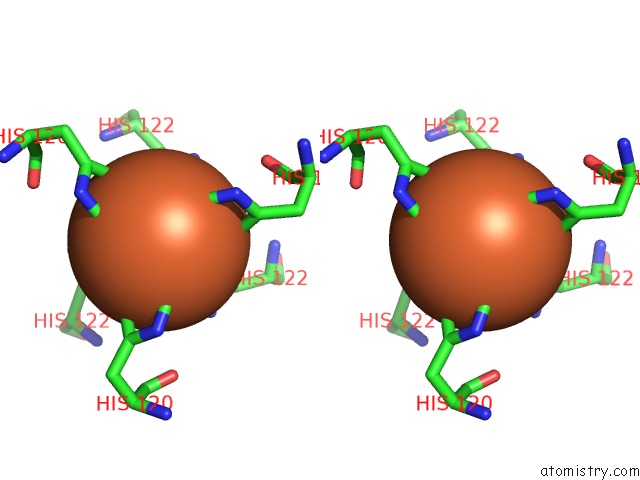
Stereo pair view
Mono view
Stereo pair view
A full contact list of Iron with other atoms in the Fe binding
site number 2 of Crystal Structure of A C-Terminal Domain of the Bacteriophage P2 Tail Spike Protein, Gpv within 5.0Å range:
|
Reference:
E.Yamashita,
A.Nakagawa,
J.Takahashi,
K.Tsunoda,
S.Yamada,
S.Takeda.
The Host-Binding Domain of the P2 Phage Tail Spike Reveals A Trimeric Iron-Binding Structure Acta Crystallogr.,Sect.F V. 67 837 2011.
ISSN: ESSN 1744-3091
PubMed: 21821878
DOI: 10.1107/S1744309111005999
Page generated: Sun Aug 4 07:19:56 2024
ISSN: ESSN 1744-3091
PubMed: 21821878
DOI: 10.1107/S1744309111005999
Last articles
Fe in 2YXOFe in 2YRS
Fe in 2YXC
Fe in 2YNM
Fe in 2YVJ
Fe in 2YP1
Fe in 2YU2
Fe in 2YU1
Fe in 2YQB
Fe in 2YOO