Iron »
PDB 3ark-3b99 »
3b6h »
Iron in PDB 3b6h: Crystal Structure of Human Prostacyclin Synthase in Complex with Inhibitor Minoxidil
Enzymatic activity of Crystal Structure of Human Prostacyclin Synthase in Complex with Inhibitor Minoxidil
All present enzymatic activity of Crystal Structure of Human Prostacyclin Synthase in Complex with Inhibitor Minoxidil:
5.3.99.4;
5.3.99.4;
Protein crystallography data
The structure of Crystal Structure of Human Prostacyclin Synthase in Complex with Inhibitor Minoxidil, PDB code: 3b6h
was solved by
Y.-C.Li,
C.-W.Chiang,
H.-C.Yeh,
P.-Y.Hsu,
F.G.Whitby,
L.-H.Wang,
N.-L.Chan,
with X-Ray Crystallography technique. A brief refinement statistics is given in the table below:
| Resolution Low / High (Å) | 29.09 / 1.62 |
| Space group | P 1 21 1 |
| Cell size a, b, c (Å), α, β, γ (°) | 68.762, 106.072, 73.901, 90.00, 91.81, 90.00 |
| R / Rfree (%) | 20.3 / 22.8 |
Iron Binding Sites:
The binding sites of Iron atom in the Crystal Structure of Human Prostacyclin Synthase in Complex with Inhibitor Minoxidil
(pdb code 3b6h). This binding sites where shown within
5.0 Angstroms radius around Iron atom.
In total 2 binding sites of Iron where determined in the Crystal Structure of Human Prostacyclin Synthase in Complex with Inhibitor Minoxidil, PDB code: 3b6h:
Jump to Iron binding site number: 1; 2;
In total 2 binding sites of Iron where determined in the Crystal Structure of Human Prostacyclin Synthase in Complex with Inhibitor Minoxidil, PDB code: 3b6h:
Jump to Iron binding site number: 1; 2;
Iron binding site 1 out of 2 in 3b6h
Go back to
Iron binding site 1 out
of 2 in the Crystal Structure of Human Prostacyclin Synthase in Complex with Inhibitor Minoxidil
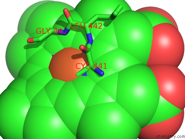
Mono view
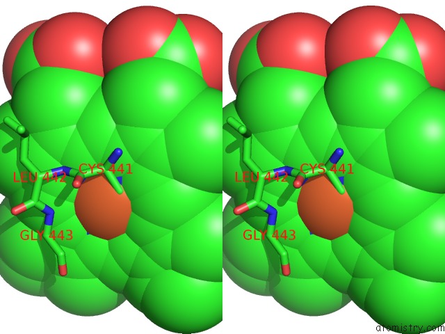
Stereo pair view
Mono view
Stereo pair view
A full contact list of Iron with other atoms in the Fe binding
site number 1 of Crystal Structure of Human Prostacyclin Synthase in Complex with Inhibitor Minoxidil within 5.0Å range:
|
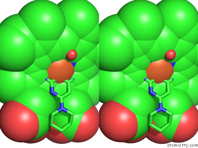
Iron binding site 2 out of 2 in 3b6h
Go back to
Iron binding site 2 out
of 2 in the Crystal Structure of Human Prostacyclin Synthase in Complex with Inhibitor Minoxidil
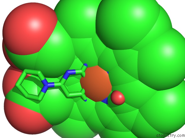
Mono view
Stereo pair view
Mono view
Stereo pair view
A full contact list of Iron with other atoms in the Fe binding
site number 2 of Crystal Structure of Human Prostacyclin Synthase in Complex with Inhibitor Minoxidil within 5.0Å range:
|
Reference:
Y.-C.Li,
C.-W.Chiang,
H.-C.Yeh,
P.-Y.Hsu,
F.G.Whitby,
L.-H.Wang,
N.-L.Chan.
Structures of Prostacyclin Synthase and Its Complexes with Substrate Analog and Inhibitor Reveal A Ligand-Specific Heme Conformation Change J.Biol.Chem. V. 283 2917 2008.
ISSN: ISSN 0021-9258
PubMed: 18032380
DOI: 10.1074/JBC.M707470200
Page generated: Sun Aug 4 07:39:38 2024
ISSN: ISSN 0021-9258
PubMed: 18032380
DOI: 10.1074/JBC.M707470200
Last articles
Zn in 9MJ5Zn in 9HNW
Zn in 9G0L
Zn in 9FNE
Zn in 9DZN
Zn in 9E0I
Zn in 9D32
Zn in 9DAK
Zn in 8ZXC
Zn in 8ZUF