Iron »
PDB 3b9j-3by0 »
3bu7 »
Iron in PDB 3bu7: Crystal Structure and Biochemical Characterization of Gdosp, A Gentisate 1,2-Dioxygenase From Silicibacter Pomeroyi
Enzymatic activity of Crystal Structure and Biochemical Characterization of Gdosp, A Gentisate 1,2-Dioxygenase From Silicibacter Pomeroyi
All present enzymatic activity of Crystal Structure and Biochemical Characterization of Gdosp, A Gentisate 1,2-Dioxygenase From Silicibacter Pomeroyi:
1.13.11.4;
1.13.11.4;
Protein crystallography data
The structure of Crystal Structure and Biochemical Characterization of Gdosp, A Gentisate 1,2-Dioxygenase From Silicibacter Pomeroyi, PDB code: 3bu7
was solved by
J.Chen,
M.Z.Wang,
G.Y.Zhu,
X.C.Zhang,
Z.H.Rao,
with X-Ray Crystallography technique. A brief refinement statistics is given in the table below:
| Resolution Low / High (Å) | 41.93 / 2.80 |
| Space group | H 3 2 |
| Cell size a, b, c (Å), α, β, γ (°) | 129.999, 129.999, 246.255, 90.00, 90.00, 120.00 |
| R / Rfree (%) | 21 / 24.5 |
Iron Binding Sites:
The binding sites of Iron atom in the Crystal Structure and Biochemical Characterization of Gdosp, A Gentisate 1,2-Dioxygenase From Silicibacter Pomeroyi
(pdb code 3bu7). This binding sites where shown within
5.0 Angstroms radius around Iron atom.
In total 4 binding sites of Iron where determined in the Crystal Structure and Biochemical Characterization of Gdosp, A Gentisate 1,2-Dioxygenase From Silicibacter Pomeroyi, PDB code: 3bu7:
Jump to Iron binding site number: 1; 2; 3; 4;
In total 4 binding sites of Iron where determined in the Crystal Structure and Biochemical Characterization of Gdosp, A Gentisate 1,2-Dioxygenase From Silicibacter Pomeroyi, PDB code: 3bu7:
Jump to Iron binding site number: 1; 2; 3; 4;
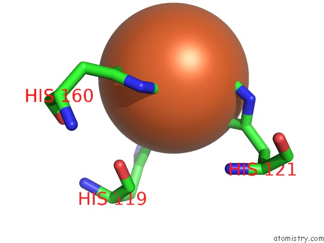
Iron binding site 1 out of 4 in 3bu7
Go back to
Iron binding site 1 out
of 4 in the Crystal Structure and Biochemical Characterization of Gdosp, A Gentisate 1,2-Dioxygenase From Silicibacter Pomeroyi
Mono view
Stereo pair view
Mono view
Stereo pair view
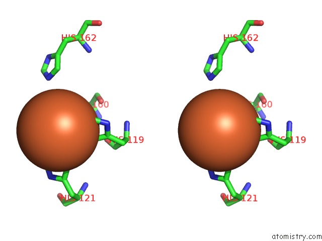
A full contact list of Iron with other atoms in the Fe binding
site number 1 of Crystal Structure and Biochemical Characterization of Gdosp, A Gentisate 1,2-Dioxygenase From Silicibacter Pomeroyi within 5.0Å range:
|
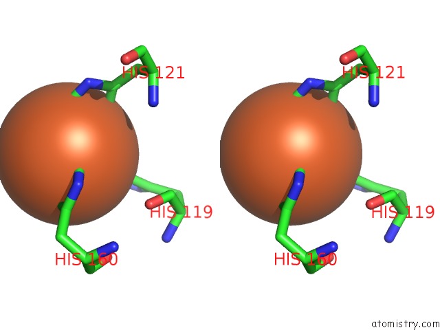
Iron binding site 2 out of 4 in 3bu7
Go back to
Iron binding site 2 out
of 4 in the Crystal Structure and Biochemical Characterization of Gdosp, A Gentisate 1,2-Dioxygenase From Silicibacter Pomeroyi

Mono view
Stereo pair view
Mono view
Stereo pair view
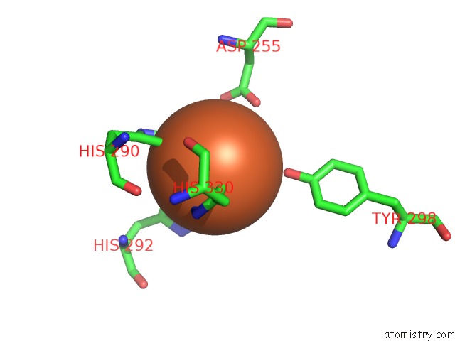
A full contact list of Iron with other atoms in the Fe binding
site number 2 of Crystal Structure and Biochemical Characterization of Gdosp, A Gentisate 1,2-Dioxygenase From Silicibacter Pomeroyi within 5.0Å range:
|
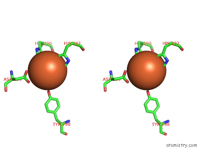
Iron binding site 3 out of 4 in 3bu7
Go back to
Iron binding site 3 out
of 4 in the Crystal Structure and Biochemical Characterization of Gdosp, A Gentisate 1,2-Dioxygenase From Silicibacter Pomeroyi
Mono view
Stereo pair view
Mono view
Stereo pair view
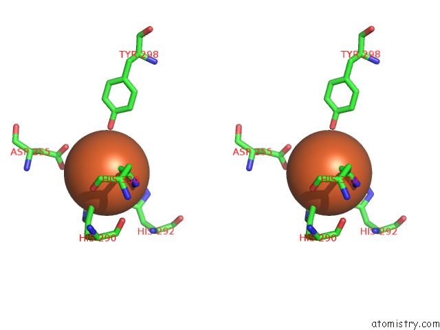
A full contact list of Iron with other atoms in the Fe binding
site number 3 of Crystal Structure and Biochemical Characterization of Gdosp, A Gentisate 1,2-Dioxygenase From Silicibacter Pomeroyi within 5.0Å range:
|
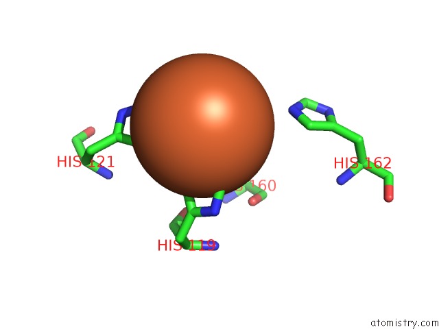
Iron binding site 4 out of 4 in 3bu7
Go back to
Iron binding site 4 out
of 4 in the Crystal Structure and Biochemical Characterization of Gdosp, A Gentisate 1,2-Dioxygenase From Silicibacter Pomeroyi
Mono view
Stereo pair view
Mono view
Stereo pair view
A full contact list of Iron with other atoms in the Fe binding
site number 4 of Crystal Structure and Biochemical Characterization of Gdosp, A Gentisate 1,2-Dioxygenase From Silicibacter Pomeroyi within 5.0Å range:
|
Reference:
J.Chen,
W.Li,
M.Wang,
G.Zhu,
D.Liu,
F.Sun,
N.Hao,
X.Li,
Z.Rao,
X.C.Zhang.
Crystal Structure and Mutagenic Analysis of Gdosp, A Gentisate 1,2-Dioxygenase From Silicibacter Pomeroyi. Protein Sci. V. 17 1362 2008.
ISSN: ISSN 0961-8368
PubMed: 18505738
DOI: 10.1110/PS.035881.108
Page generated: Mon Aug 4 23:59:48 2025
ISSN: ISSN 0961-8368
PubMed: 18505738
DOI: 10.1110/PS.035881.108
Last articles
Fe in 3IAMFe in 3I9V
Fe in 3IR5
Fe in 3IQB
Fe in 3IQ6
Fe in 3IQ5
Fe in 3IIZ
Fe in 3IIX
Fe in 3ICF
Fe in 3IC2