Iron »
PDB 3b9j-3by0 »
3buj »
Iron in PDB 3buj: Crystal Structure of CALO2
Protein crystallography data
The structure of Crystal Structure of CALO2, PDB code: 3buj
was solved by
J.G.Mccoy,
H.D.Johnson,
S.Singh,
C.A.Bingman,
J.S.Thorson,
G.N.Phillipsjr.,
with X-Ray Crystallography technique. A brief refinement statistics is given in the table below:
| Resolution Low / High (Å) | 67.27 / 2.47 |
| Space group | P 41 21 2 |
| Cell size a, b, c (Å), α, β, γ (°) | 71.202, 71.202, 204.451, 90.00, 90.00, 90.00 |
| R / Rfree (%) | 19.8 / 25.7 |
Iron Binding Sites:
The binding sites of Iron atom in the Crystal Structure of CALO2
(pdb code 3buj). This binding sites where shown within
5.0 Angstroms radius around Iron atom.
In total only one binding site of Iron was determined in the Crystal Structure of CALO2, PDB code: 3buj:
In total only one binding site of Iron was determined in the Crystal Structure of CALO2, PDB code: 3buj:
Iron binding site 1 out of 1 in 3buj
Go back to
Iron binding site 1 out
of 1 in the Crystal Structure of CALO2

Mono view
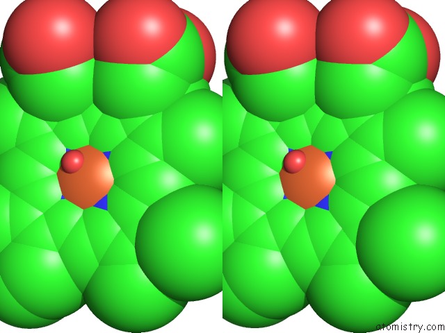
Stereo pair view
Mono view
Stereo pair view
A full contact list of Iron with other atoms in the Fe binding
site number 1 of Crystal Structure of CALO2 within 5.0Å range:
|
Reference:
J.G.Mccoy,
H.D.Johnson,
S.Singh,
C.A.Bingman,
I.K.Lei,
J.S.Thorson,
G.N.Phillips Jr..
Structural Characterization of CALO2: A Putative Orsellinic Acid P450 Oxidase in the Calicheamicin Biosynthetic Pathway. Proteins V. 74 50 2009.
ISSN: ISSN 0887-3585
PubMed: 18561189
DOI: 10.1002/PROT.22131
Page generated: Sun Aug 4 08:03:16 2024
ISSN: ISSN 0887-3585
PubMed: 18561189
DOI: 10.1002/PROT.22131
Last articles
Zn in 9J0NZn in 9J0O
Zn in 9J0P
Zn in 9FJX
Zn in 9EKB
Zn in 9C0F
Zn in 9CAH
Zn in 9CH0
Zn in 9CH3
Zn in 9CH1